Fluid Pressure and Cell Changes Drive Blood Vessel Growth

Greg Howard
23rd February, 2025

Fluid Pressure and Cell Changes Drive Blood Vessel Growth

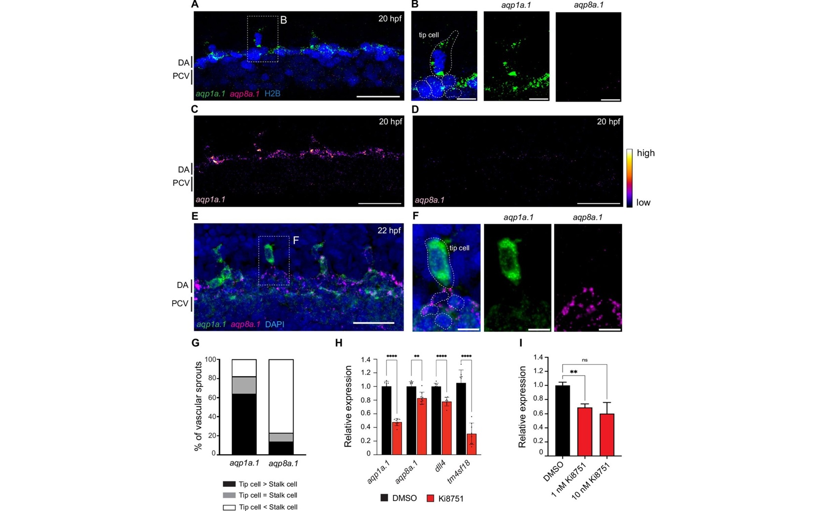
High-resolution imaging of vascular sprouts in Zebrafish (Danio rerio) reveals differential enrichment of aqp1a.1 in tip cells and aqp8a.1 in stalk cells (a–g), while quantitative PCR assays demonstrate that the expression of these water channels is induced by VEGFR2 signaling in both zebrafish and human endothelial cells (h, i).

Image adapted from: Kondrychyn et al. / CC BY (Source)

Key Findings

  • *Researchers in Kobe, Japan discovered that Aquaporin proteins help blood vessel cells move even when usual movement methods are blocked.*
  • *Disabling these Aquaporins in zebrafish leads to slower and impaired blood vessel formation due to smaller cell size and fewer membrane extensions.*
  • *Manipulating Aquaporin activity could offer new ways to promote or inhibit blood vessel growth in various diseases.*
Cell migration is essential for the development and maintenance of tissues in living organisms. Understanding the mechanisms behind cell movement can provide insights into various biological processes and diseases. A recent study conducted by researchers at RIKEN in Kobe, Japan, sheds light on a novel mechanism that endothelial cells (ECs) use to migrate and form new blood vessels, a process known as sprouting angiogenesis[1]. Traditionally, cell migration has been associated with the actomyosin network, a structure composed of actin filaments and myosin motors that generates contractile forces allowing cells to move. However, the RIKEN study discovered that endothelial tip cells, which lead the formation of new blood vessels, can still migrate even when actin polymerization is inhibited. This unexpected finding suggested the presence of an alternative migration mechanism. The researchers found that endothelial tip cells rely on Aquaporins (Aqp), specifically Aqp1a.1 and Aqp8a.1, to facilitate water inflow into the cells. Aquaporins are membrane proteins that form channels allowing water to pass through cell membranes. By increasing the flow of water into the cells, these proteins help raise the hydrostatic pressure within the cells, leading to an expansion in cell volume. This increase in pressure generates membrane protrusions, which are essential for the cells to move forward and form new blood vessels. In their experiments using zebrafish, a common model organism for studying vascular development, the researchers observed that the expression of aqp1a.1 and aqp8a.1 was regulated by VEGFR2, a receptor involved in blood vessel formation. When the function of these aquaporins was disrupted, the formation of intersegmental vessels was impaired. The endothelial tip cells were less capable of increasing their cytoplasmic volume and generating the necessary membrane protrusions, resulting in delayed emergence from the dorsal aorta and slower overall migration. Furthermore, when actin polymerization was also inhibited, the sprouting angiogenesis was significantly more affected, indicating that ECs utilize both the traditional actomyosin-dependent and the newly discovered aquaporin-mediated mechanisms to ensure robust cell migration. This study builds upon previous research that highlights the importance of fluid dynamics and hydrostatic pressure in tissue development. For instance, a study on mouse blastocysts revealed that fluid-filled lumens generate pressure that influences embryo size and cell fate by regulating tissue mechanics[2]. Similarly, research on diabetic microvascular complications demonstrated that aquaporins play a critical role in maintaining blood vessel diameter and perfusion, with their dysfunction leading to reduced vascular diameter and insufficient blood supply[3]. Additionally, studies have shown that active fluid transport across epithelial layers acts as a driving force for tissue morphogenesis, emphasizing the role of hydraulic pressure in shaping tissues[4][5]. By integrating these insights, the RIKEN study not only identifies a new migration mechanism but also connects it to the broader understanding of how fluid dynamics influence tissue development and vascular health. The finding that aquaporins contribute to endothelial cell migration through hydrostatic pressure adds a crucial piece to the puzzle of how tissues grow and maintain their structure. This dual mechanism of cell migration ensures that blood vessels can form efficiently even under conditions where traditional actin-dependent forces are compromised. Furthermore, the study's discovery has potential implications for therapeutic strategies. For example, enhancing the function of aquaporins could be a promising approach to promote blood vessel formation in diseases where vascular growth is needed, such as in wound healing or in treating certain cardiovascular conditions. Conversely, targeting aquaporins might help inhibit unwanted blood vessel growth in diseases like cancer, where excessive angiogenesis can facilitate tumor growth and metastasis. The use of zebrafish as a model organism was particularly advantageous in this research due to their transparent embryos and rapid development, which allow for real-time observation of vascular formation and cell migration. This model system enabled the researchers to manipulate gene function and monitor the resulting effects on blood vessel development directly. In conclusion, the study from RIKEN advances our understanding of endothelial cell migration by revealing the significant role of aquaporin-mediated water inflow and hydrostatic pressure. This mechanism operates alongside the well-established actomyosin-dependent forces, providing a more comprehensive picture of the forces driving tissue morphogenesis. By linking fluid dynamics to cellular behavior, this research opens new avenues for exploring how mechanical and molecular factors collaborate to shape living tissues, offering potential targets for therapeutic intervention in various vascular diseases.

MedicineHealthBiochem

References

Main Study

1) Combined forces of hydrostatic pressure and actin polymerization drive endothelial tip cell migration and sprouting angiogenesis

Published 20th February, 2025

https://doi.org/10.7554/eLife.98612


Related Studies

2) Hydraulic control of mammalian embryo size and cell fate.

https://doi.org/10.1038/s41586-019-1309-x


3) Aquaporins enriched in endothelial vacuole membrane regulate the diameters of microvasculature in hyperglycaemia.

https://doi.org/10.1093/cvr/cvae085


4) Trans-epithelial fluid flow and mechanics of epithelial morphogenesis.

https://doi.org/10.1016/j.semcdb.2022.05.020


5) Hydrostatic pressure as a driver of cell and tissue morphogenesis.

https://doi.org/10.1016/j.semcdb.2022.04.021


Related Articles

An unhandled error has occurred. Reload 🗙