Computer vision speeds up research on two-spotted spider mite development

Jenn Hoskins
30th December, 2025

Computer vision speeds up research on two-spotted spider mite development

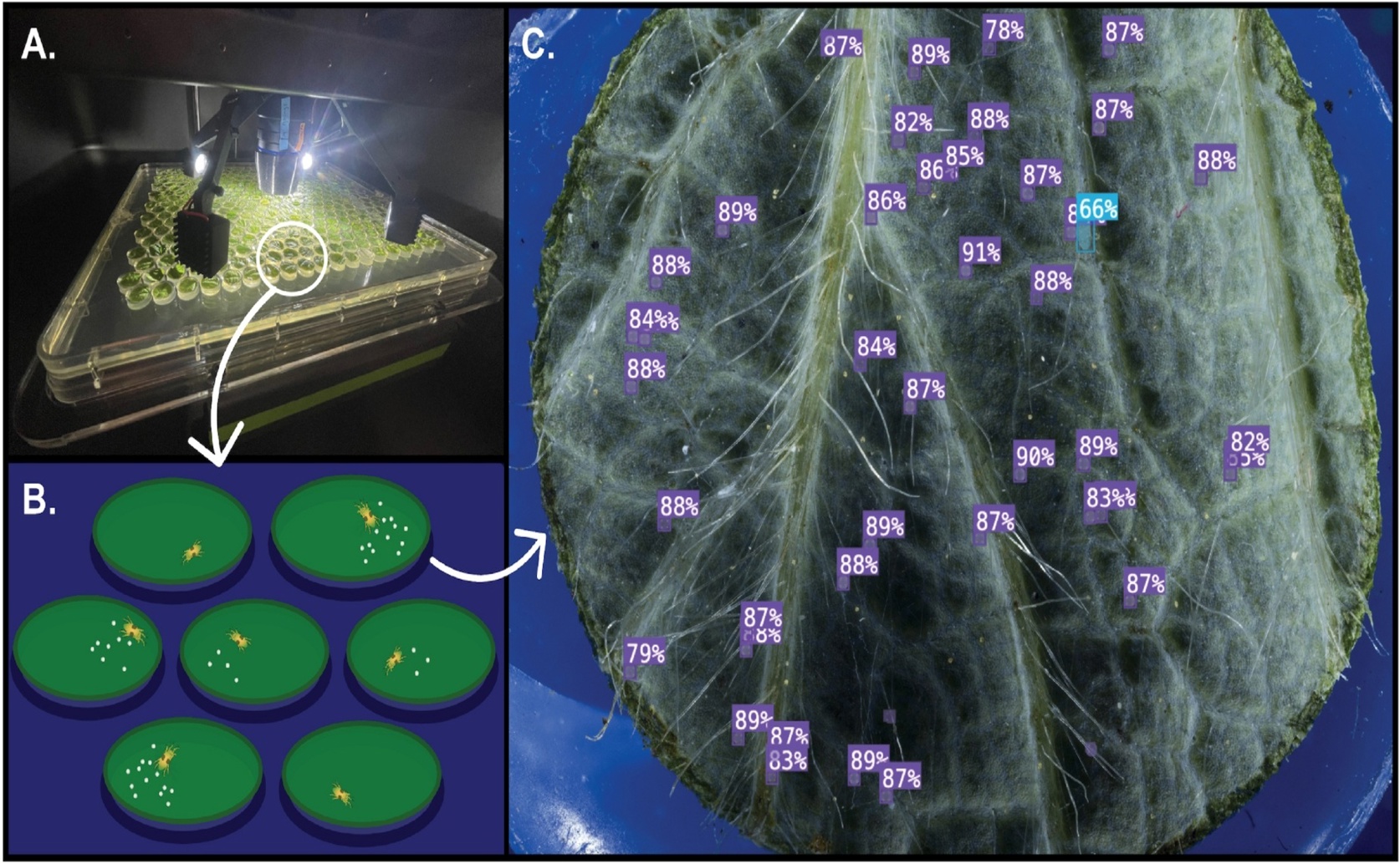
The high-throughput phenotyping pipeline integrates the Blackbird automated imaging platform (a) with a standardized in vitro assay (b), enabling a computer vision model to successfully detect and classify different life stages of the two-spotted spider mite (Tetranychus urticae) on a leaf disk (c).

Image adapted from: Wiseman et al. / CC BY (Source)

Key Findings

  • Researchers developed a new automated system to quickly count spider mites on plants, addressing a bottleneck in breeding pest-resistant crops
  • The system uses computer vision models trained on a large dataset of over 32,000 mite images to accurately identify mite life stages, achieving high precision
  • The automated system effectively measured mite reproduction rates, closely matching manual counts, and offers a faster, more standardized way to screen for pest resistance
The two-spotted spider mite (Tetranychus urticae) is a pervasive agricultural pest, known for its quick reproduction, fast development, and ability to rapidly evolve resistance to pesticides, known as miticides. Traditional methods for breeding crops resistant to these mites are effective, but are often slow and require significant manual effort to identify resistant plants. This limits the widespread use of resistant cultivars as a sustainable pest control strategy. Researchers at Oregon State University, in collaboration with the United States Department of Agriculture and Guizhou University, have developed a new automated system to speed up this process[1]. The core problem this research addresses is the bottleneck in “phenotyping” spider mites – accurately assessing their life stages (egg, larvae, nymph, adult) and reproductive rates on different plants. Traditionally, this is done by visually counting mites under a microscope, a process that is time-consuming and prone to human error. The new system, detailed in, utilizes a high-throughput imaging pipeline. This involves a robotic microscope (Blackbird CNC Microscopy Imaging Robot) combined with computer vision models to automatically identify and count mites in images. The project began by creating a large, publicly available dataset of over 1,500 annotated images, containing nearly 32,000 individual mites labeled by life stage. These images spanned five different categories of biological relevance, across ten host plant species and over 25 different cultivars (varieties) of those plants. This diverse dataset was crucial for training the computer vision models. The researchers tested three different versions of a machine learning model called YOLO11, each configured to identify a different number of life stages – three, four, or five classes. These models were “trained” using both real images from the dataset and synthetically generated images to improve their accuracy. The three-class model, distinguishing eggs, juveniles and adults, performed the best overall, achieving high precision (0.875), recall (0.871), and mean Average Precision (mAP50 = 0.883). Precision refers to the accuracy of positive identifications (correctly identifying mites when they are present), while recall measures the ability to find all mites present in an image. mAP50 is a combined metric representing overall accuracy. Importantly, the model was robust to variations in the background (different host plants) and worked well even when there were moderate numbers of mites in the image. To validate the system, the researchers applied it to standard miticide resistance assays. The system accurately estimated the reproductive rate (fecundity) of the mites, but was less accurate at determining mortality rates, sometimes misclassifying dead mites as alive. This highlights a limitation of the system – accurately identifying dead mites is more challenging. Further testing on hop cultivars showed the pipeline could reliably detect differences in fecundity, closely matching results obtained through manual counting (R2≥ 0.98). The study also identified limitations. Performance decreased when the system was used on host plants not included in the training dataset, emphasizing the need for “fine-tuning” the models with data specific to each host. Similarly, accuracy declined at high mite densities (more than 80 mites per image), suggesting the need for experimental designs that avoid overcrowding. Understanding the digestive and detoxification capabilities of spider mites is critical for developing effective resistance breeding strategies[2][3]. The genome of T. urticae reveals an expansion of gene families involved in these processes, linked to the mite’s ability to adapt to different host plants and overcome plant defenses. The research in provides a tool to more efficiently screen for traits related to these adaptations. By enabling faster and more standardized quantification of mite life stages, this automated system offers a scalable alternative to manual scoring, particularly for resistance breeding programs focused on antibiosis – the ability of a plant to reduce mite reproduction or survival. The dataset, code, and trained models are publicly available, allowing other researchers to adopt and extend this technology.

AgricultureBiotechPlant Science

References

Main Study

1) Automated detection and quantification of two-spotted spider mite life stages using computer vision for high-throughput in vitro assays

Published 29th December, 2025

https://doi.org/10.1371/journal.pone.0333253


Related Studies

2) The Digestive System of the Two-Spotted Spider Mite, Tetranychus urticae Koch, in the Context of the Mite-Plant Interaction.

https://doi.org/10.3389/fpls.2018.01206


3) The genome of Tetranychus urticae reveals herbivorous pest adaptations.

https://doi.org/10.1038/nature10640


Related Articles

An unhandled error has occurred. Reload 🗙