AI Tool Finds and Separates Kidney Parts in Fluorescent Images

Jenn Hoskins
15th April, 2025

AI Tool Finds and Separates Kidney Parts in Fluorescent Images

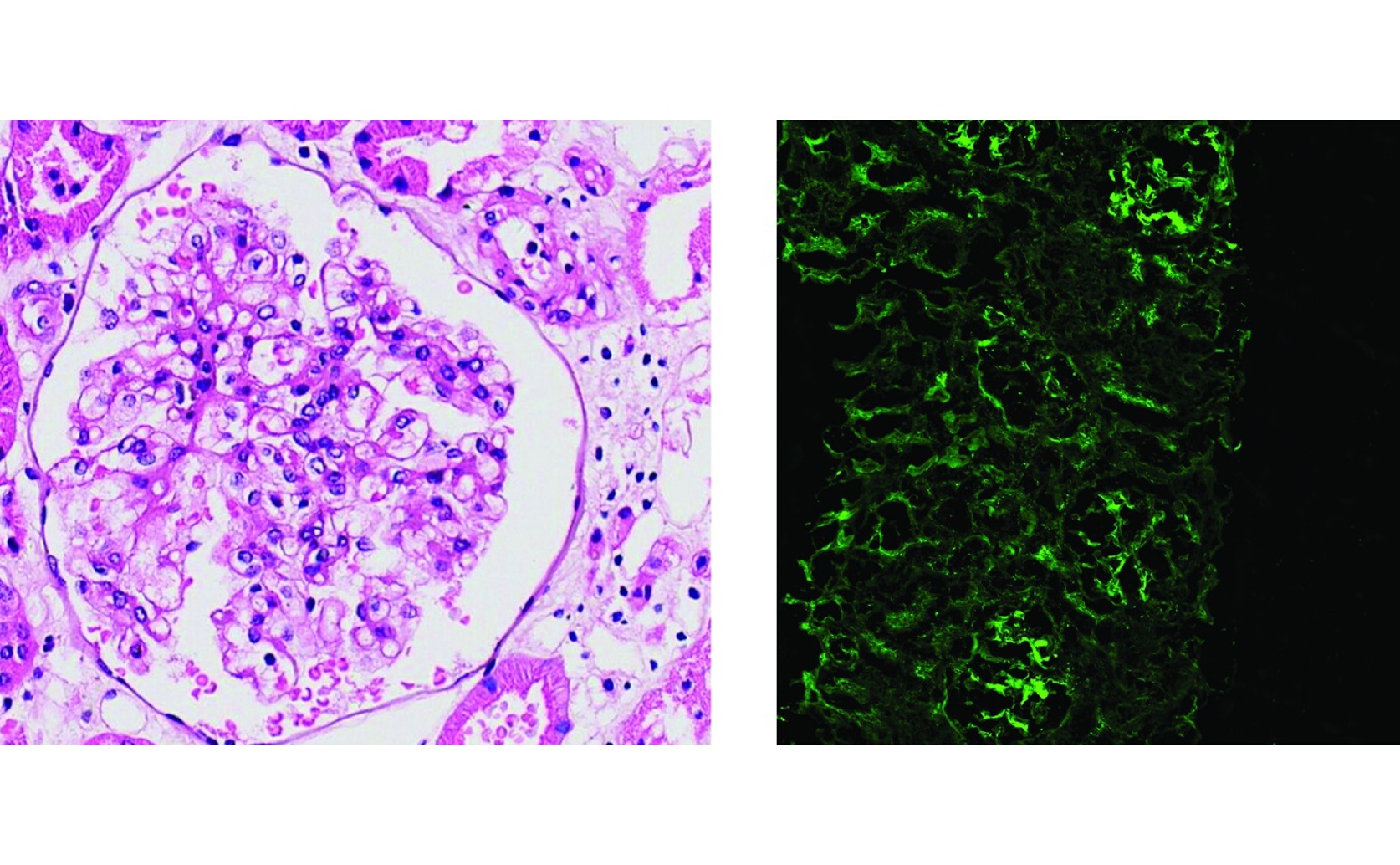
This figure contrasts a PAS-stained image (left) with an immunofluorescence (IF) image (right) to highlight the study's central challenge: the indistinct boundaries of glomeruli in IF images make them significantly more difficult to segment.

Image adapted from: Pan et al. / CC BY (Source)

Key Findings

  • Researchers at South China University of Technology created GlomSAM, a new tool that accurately identifies kidney's filtering units in medical images
  • GlomSAM outperforms existing methods by over 15%, making early detection of chronic kidney disease more reliable
  • This innovation reduces the need for manual work, speeding up diagnosis and improving consistency in kidney disease detection
Chronic kidney disease (CKD) often progresses silently, making early detection and diagnosis crucial for effective management. A key aspect of diagnosing CKD involves analyzing glomeruli, the tiny filtering units within the kidneys. Accurate segmentation of these structures in immunofluorescence images is essential but traditionally relies on labor-intensive manual annotations. Existing automated methods struggle with low recall rates and limited accuracy, hindering their practical utility. Researchers at the South China University of Technology have introduced a novel approach to address these challenges through a SAM-based multi-glomerular segmentation model, named GlomSAM[1]. This study leverages the recently developed Segment Anything Model (SAM), which has shown promising results in various segmentation tasks across different domains. By adapting SAM to the specific needs of immunofluorescence pathology, GlomSAM aims to enhance the accuracy and efficiency of glomerular segmentation. To optimize SAM for pathological analysis, the researchers employed a fusion encoder strategy that combines the strengths of convolutional neural networks (CNNs) and transformer architectures. This hybrid approach facilitates better feature extraction and supports effective transfer learning, enabling the model to generalize well across different pathological images. Additionally, GlomSAM incorporates a rough mask generator that creates preliminary segmentation masks. These masks serve as automated input prompts, refining the final segmentation results and reducing the dependency on manual annotations. The effectiveness of GlomSAM was rigorously tested through extensive comparative experiments and ablation studies. The results demonstrated that GlomSAM achieved state-of-the-art performance, outperforming existing segmentation methods in terms of both recall rates and overall accuracy. This advancement builds upon previous efforts to improve glomerular segmentation. For instance, a study from Zhejiang Chinese Medical University introduced an unsupervised stain augmentation method[2], which enhanced segmentation performance by increasing staining diversity and employing advanced feature extraction techniques. Similarly, research by Ningbo KonFoong Bioinformation Tech Co. Ltd developed CNN-based models for identifying and classifying glomerular features, achieving high F1-scores and showing agreement with pathologist assessments[3]. Furthermore, the Glo-In-One toolkit developed by South China University of Technology provided a user-friendly solution for glomerular detection and segmentation, along with a large dataset to facilitate further algorithmic development[4]. GlomSAM advances the field by integrating these previous innovations into a cohesive model tailored for immunofluorescence images. Unlike traditional methods that require extensive manual labeling, GlomSAM's automated prompting and refined segmentation capabilities significantly reduce the need for labor-intensive annotations. This not only speeds up the diagnostic process but also enhances the consistency and reliability of glomerular segmentation. The implications of this study are substantial for both clinical practice and research. By enabling more accurate and efficient glomerular segmentation, GlomSAM can support earlier detection of CKD, potentially improving patient outcomes through timely intervention. Additionally, the model's ability to generalize across different staining methods and imaging conditions makes it a versatile tool for various applications in digital renal pathology. Looking ahead, the success of GlomSAM opens avenues for further research and development. Future studies could explore the integration of additional data modalities or the application of similar models to other complex segmentation tasks within pathology. Moreover, combining GlomSAM with existing toolkits like Glo-In-One could enhance the overall workflow for nephrologists and researchers, providing a more comprehensive suite of tools for kidney disease analysis. In conclusion, the development of GlomSAM represents a significant step forward in the automated analysis of immunofluorescence images for chronic kidney disease. By building on previous advancements and introducing innovative strategies for segmentation, this study offers a robust solution to a longstanding challenge in nephrology research. The continued evolution of such models holds promise for improving diagnostic accuracy and efficiency, ultimately contributing to better patient care and advancing our understanding of kidney diseases.

MedicineBiotechBiochem

References

Main Study

1) GlomSAM: Hybrid customized SAM for multi-glomerular detection and segmentation in immunofluorescence images

Published 14th April, 2025

https://doi.org/10.1371/journal.pone.0321096


Related Studies

2) Unsupervised stain augmentation enhanced glomerular instance segmentation on pathology images.

https://doi.org/10.1007/s11548-024-03154-7


3) Artificial intelligence assists identification and pathologic classification of glomerular lesions in patients with diabetic nephropathy.

https://doi.org/10.1186/s12967-024-05221-8


4) Glo-In-One: holistic glomerular detection, segmentation, and lesion characterization with large-scale web image mining.

https://doi.org/10.1117/1.JMI.9.5.052408


Related Articles

An unhandled error has occurred. Reload 🗙