Natural Copper Oxide Particles as Cancer and Bacteria Fighters and How They Work

Jenn Hoskins
4th April, 2025

Natural Copper Oxide Particles as Cancer and Bacteria Fighters and How They Work

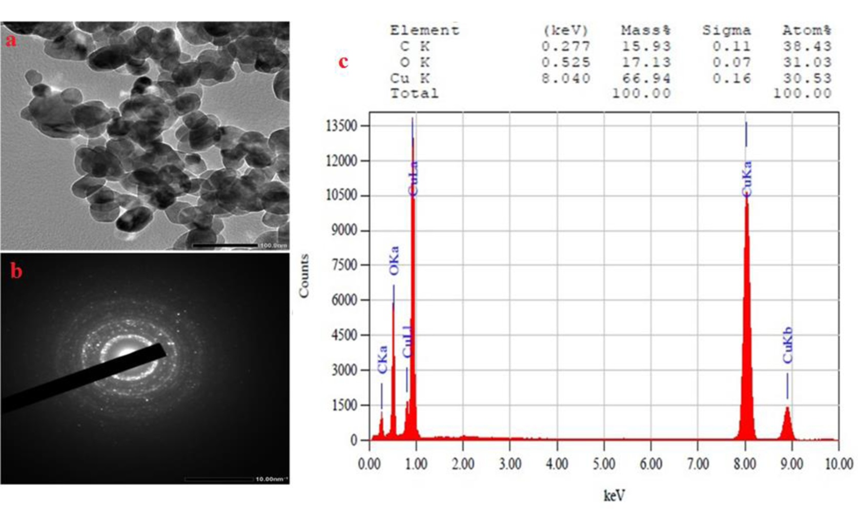
Morphological and elemental analyses confirm the successful green synthesis of copper oxide nanoparticles, revealing their predominantly spherical and hexagonal shape with a size of 10-30 nm (a), crystalline structure (b), and high elemental purity (c).

Image adapted from: Gad et al. / CC BY (Source)

Key Findings

  • Researchers at Jouf University used okra extract to eco-friendly produce tiny copper oxide particles
  • These copper oxide nanoparticles safely targeted cancer cells, showing promise for cancer treatment
  • They also effectively inhibited harmful bacteria, especially Staphylococcus aureus, highlighting their antibacterial potential
Nanoparticles, tiny particles measured in nanometers, have revolutionized various scientific fields due to their unique properties. Traditional methods of producing these particles often involve chemical and physical processes that can be harmful to the environment. However, green synthesis offers a sustainable alternative by utilizing plant extracts to create nanoparticles in an eco-friendly manner. A recent study by researchers at Jouf University[1] explored this approach by using okra fruit extract to produce copper oxide nanoparticles (CuO NPs), highlighting their potential applications in antibacterial and anticancer treatments. The green synthesis method employed by Jouf University is both simple and cost-effective. By using okra extract, the researchers took advantage of the plant's natural phytochemicals, which act as capping and stabilizing agents, similar to the processes described in earlier research[2]. This method not only reduces the environmental impact associated with conventional nanoparticle production but also ensures that the resulting CuO NPs are highly stable. To confirm the successful synthesis of CuO NPs, the team utilized several analytical techniques. UV-visible spectroscopy revealed a maximum absorbance at 381 nm, while Fourier-Transform Infrared Spectroscopy (FT-IR) identified characteristic bands at 534 and 588 cm⁻¹. X-Ray Diffraction (XRD) analysis confirmed the monoclinic structure of the nanoparticles, and Transmission Electron Microscopy (TEM) showed that the particles ranged in size from 10 to 30 nm. Additionally, Energy-Dispersive X-ray (EDX) analysis provided further confirmation of the elemental composition. These detailed characterizations align with findings from related studies that emphasize the importance of using multiple techniques to ensure the accuracy of nanoparticle synthesis[2]. The study also assessed the biological activities of the synthesized CuO NPs. In cytotoxicity tests conducted on Human Fibroblast normal HFB4 cell lines, the nanoparticles exhibited a half-maximal inhibitory concentration (IC50) of 236.34 μg/mL, indicating a moderate level of safety. More notably, the CuO NPs demonstrated significant antitumor effects against breast adenocarcinoma Mcf-7 cell lines, with an IC50 of 109.46 μg/mL. This suggests that CuO NPs could be effective in targeting cancer cells while maintaining a degree of safety for normal cells, a balance that is crucial in cancer therapy. The antibacterial properties of CuO NPs were also thoroughly investigated. The nanoparticles showed substantial inhibition zones against Staphylococcus aureus and Bacillus cereus, measuring 29.5 ± 0.7 mm and 24.6 ± 1.2 mm, respectively. In contrast, Pseudomonas aeruginosa was less affected, with an inhibition zone of 15 ± 1.6 mm. This selective antibacterial activity is consistent with previous studies that have found copper-based nanoparticles to be more effective against Gram-positive bacteria compared to Gram-negative strains[2]. The enhanced effectiveness against Gram-positive bacteria can be attributed to differences in their cell wall structures, which impact how nanoparticles interact with them. Furthermore, molecular docking studies revealed that CuO NPs interact with the dihydrofolate reductase (DHFR) enzyme of Staphylococcus aureus. The nanoparticles partially interlocked with the active site of DHFR through five classical hydrogen bonds, similar to the binding mechanism of the antifolate drug methotrexate (MTX). This interaction suggests that CuO NPs could inhibit bacterial growth by disrupting essential enzymatic processes, adding a potential mechanism to their antibacterial action. Comparing these findings with previous research on other metal nanoparticles, such as silver and zinc oxide, provides a broader context for the significance of this study. For instance, silver nanoparticles synthesized using catfish epidermal mucus proteins demonstrated strong antimicrobial and anticancer activities[3]. Similarly, zinc oxide nanoparticles produced from Cassia javanica showed notable antibacterial, antioxidant, and antiviral properties[4]. The CuO NPs developed by Jouf University not only match but also expand upon these capabilities, offering a versatile option for biomedical applications. The integration of green synthesis methods with the potent biological activities of CuO NPs exemplifies the advancements in sustainable nanotechnology. By leveraging plant extracts, researchers can produce nanoparticles that are not only effective in combating bacterial infections and cancer cells but also environmentally benign. This approach aligns with the growing emphasis on sustainable practices in scientific research and industrial applications. In conclusion, the study conducted by Jouf University demonstrates the potential of okra-mediated green synthesis in producing copper oxide nanoparticles with significant antibacterial and anticancer properties. By building on previous research and utilizing environmentally friendly methods, this work paves the way for the development of safe and effective nanoparticle-based therapies.

MedicineBiotechBiochem

References

Main Study

1) A comprehensive study on characterization of biosynthesized copper-oxide nanoparticles, their capabilities as anticancer and antibacterial agents, and predicting optimal docking poses into the cavity of S. aureus DHFR

Published 1st April, 2025

https://doi.org/10.1371/journal.pone.0319791


Related Studies

2) Current status of plant metabolite-based fabrication of copper/copper oxide nanoparticles and their applications: a review.

https://doi.org/10.1186/s40824-020-00188-1


3) Hindering the biofilm of microbial pathogens and cancer cell lines development using silver nanoparticles synthesized by epidermal mucus proteins from Clarias gariepinus.

https://doi.org/10.1186/s12896-024-00852-7


4) Investigating the in vitro antibacterial, antibiofilm, antioxidant, anticancer and antiviral activities of zinc oxide nanoparticles biofabricated from Cassia javanica.

https://doi.org/10.1371/journal.pone.0310927


Related Articles

An unhandled error has occurred. Reload 🗙