How Fruit Fly Cells Build Their Outer Shells Using Different Methods
Greg Howard
9th September, 2025


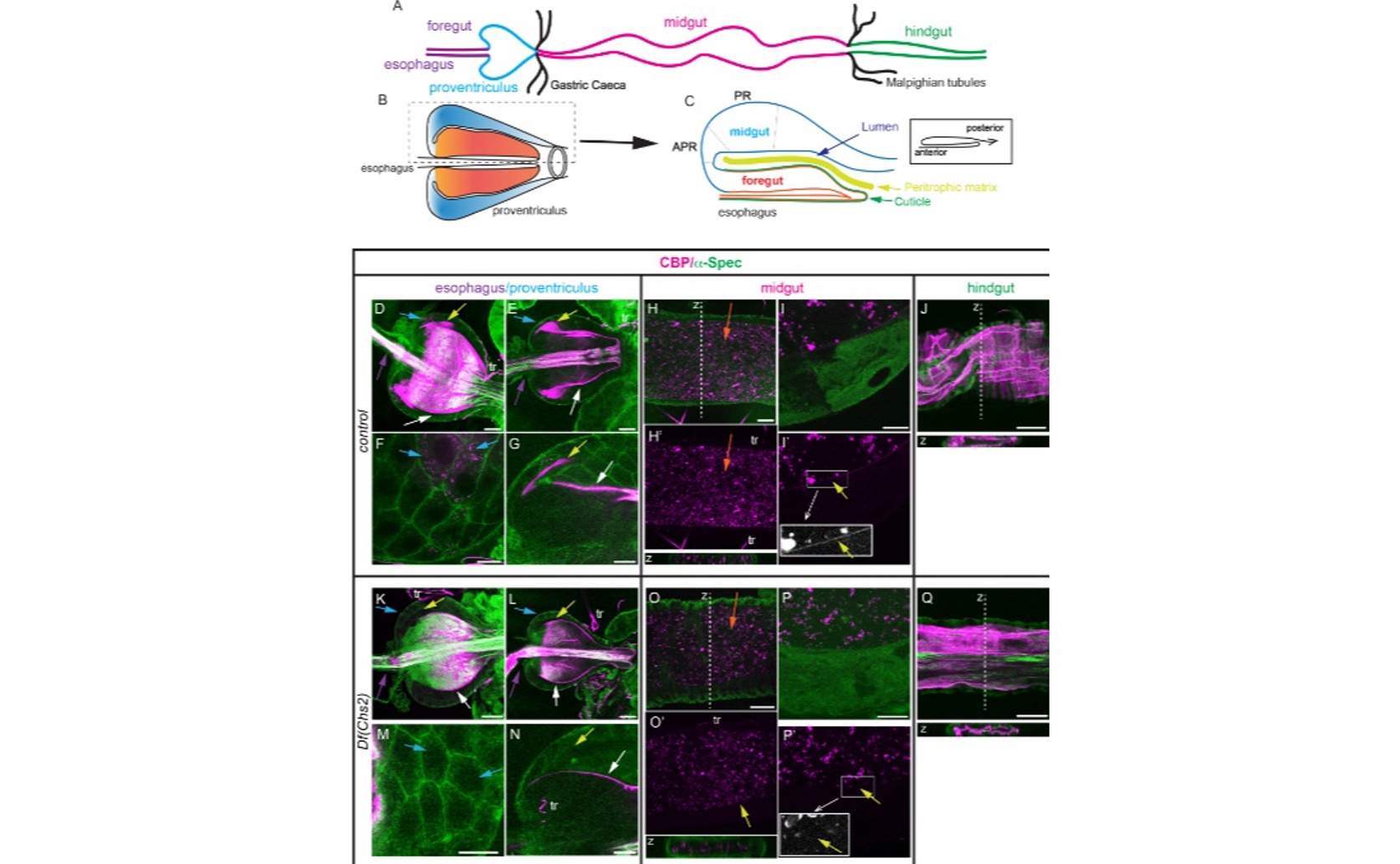
Fluorescence imaging of the Drosophila melanogaster larval digestive tract (a–c) identifies Chs2 as the enzyme responsible for synthesizing the peritrophic matrix, evidenced by the specific loss of chitin in the proventriculus and midgut of Chs2-deficient mutants (k–p) compared to controls (d–i), while ectodermal chitin deposition in the hindgut remains unaffected (j, q).
Key Findings
- In fruit flies, two chitin synthase types, Kkv and Chs2, are specifically active in different tissues: Kkv in the exoskeleton and Chs2 in the gut’s peritrophic matrix
- Kkv and Chs2 are not interchangeable, as expressing one in the other’s tissue fails to produce functional exoskeleton or peritrophic matrix structures
- Chs2 and Kkv rely on different auxiliary proteins for their function, suggesting distinct mechanisms for chitin deposition despite both being involved in chitin synthesis
References
Main Study
1) Distinct cellular and molecular mechanisms contribute to the specificity of the two Drosophila melanogaster chitin synthases in chitin deposition
Published 8th September, 2025
https://doi.org/10.1371/journal.pgen.1011847
Related Studies
2) Chitin: Structure, Chemistry and Biology.
3) Advances in understanding insect chitin biosynthesis.
4) Biosynthesis, Turnover, and Functions of Chitin in Insects.
Related Articles
 8th April, 2025 | Greg Howard