How Our Brain Starts Forming

Greg Howard
7th July, 2025

How Our Brain Starts Forming

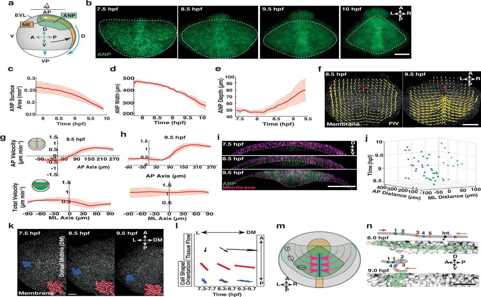
Quantitative analysis of the zebrafish (Danio rerio) anterior neural plate demonstrates that global tissue flows (f–h) drive dramatic tissue reshaping and thickening (a–e) through a coordinated process of sequential cell internalisation and multilayer folding along the dorsal midline (i–n).

Image adapted from: Inman et al. / CC BY (Source)

Key Findings

  • A University of Warwick study on zebrafish embryos reveals that the developing brain's shape is actively sculpted by physical forces, not solely by genetic instructions
  • These forces, generated by migrating cells and cell-to-cell stickiness (E-cadherin), create tissue flows that cause brain precursor cells to move inward and fold
  • This inward movement and folding are crucial for initial brain shaping, while a separate process called convergent extension is needed for the brain tissue to lengthen
The intricate process by which a single fertilized cell develops into a complex organism with distinct shapes and organs has long fascinated scientists. For centuries, the prevailing view was that an organism's final form, or its morphology, was almost entirely dictated by its genetic blueprint – the genes encoded in its DNA[2]. This perspective suggested a largely deterministic program, where genes provided all the instructions for building tissues and organs. However, recent scientific advancements have begun to reveal a more nuanced picture, showing that while genes are undoubtedly fundamental, they are not the sole determinants of shape. Instead, the physical forces and mechanical properties of cells and tissues play an equally crucial role, acting as a dynamic source of information that guides development[3][4]. Understanding how these mechanical forces contribute to shaping tissues has been a significant challenge. A new study from the University of Warwick[1] has shed light on this complex interplay by investigating the formation of the anterior neural plate (ANP) in zebrafish embryos. The ANP is a vital precursor to the forebrain, the part of the brain responsible for higher-level functions, and its proper shaping is critical for healthy brain development. The researchers aimed to uncover the specific mechanical mechanisms that regulate the ANP's shape during gastrulation, an early embryonic stage where cells rearrange to form the basic body plan. The study reveals that the ANP's shape is not simply laid down by genetic instructions but is actively sculpted by large-scale movements of tissue, referred to as global tissue flows. These flows are generated by distinct force-producing processes acting within and between different tissues. One key process identified is the migration of the mesendoderm, a group of cells that will eventually form internal organs and muscles. As these mesendoderm cells move, they exert forces on the overlying neuroectoderm, the tissue that forms the nervous system, including the ANP. Another critical factor is the interaction between different tissues, specifically mediated by a protein called E-cadherin. E-cadherin is a type of adhesion molecule found on the surface of cells, helping them stick together and communicate. The study found that E-cadherin-dependent differential tissue interactions – meaning varying strengths of adhesion between different cell types – control distinct patterns of tissue flow within the neuroectoderm. This highlights how not only large-scale movements but also subtle differences in cell-to-cell stickiness contribute to the overall mechanical landscape. The researchers observed that initial opposing flows within the neuroectoderm lead to a process called cell internalisation. This is where cells move from the outer surface of the embryo to the inside, a fundamental step in forming internal structures. Following this internalisation, the tissue undergoes progressive multilayer folding. Think of it like a sheet of paper being pushed from opposite ends, causing it to buckle and fold upon itself. These folding events, driven by the mechanical stresses from the opposing flows, in turn provide the forces necessary to reshape the ANP tissue. The study also investigated the role of convergent extension, a well-known morphogenetic process where tissues narrow in one direction while elongating in another, similar to stretching a piece of dough. They found that while convergent extension is essential for the ANP tissue to extend and lengthen, it is not required for the initial internalisation and folding events. This suggests that different mechanical mechanisms are responsible for distinct aspects of tissue shaping, operating in a coordinated but sometimes independent manner. This finding aligns with the conceptual framework proposed in earlier research, which suggests that tissue dynamics arise from a small set of cell behaviors like shape changes, cell migration, and cell contact remodeling, all requiring control over cell mechanics[3]. This research from the University of Warwick significantly advances our understanding of how complex shapes emerge during development. It provides concrete examples of how physical forces, both those generated within cells (cell-intrinsic forces, like the tension from the actomyosin network discussed in[4]) and those exerted by neighboring tissues or the environment (cell-extrinsic forces, such as the mesendoderm migration or substrate stiffness mentioned in[4]), are translated into the precise biochemical signals that guide cell behaviors and ultimately define tissue shape and size. By combining in vivo observations (studying live zebrafish embryos) with in silico modeling (computer simulations), the scientists were able to unravel the intricate spatiotemporal regulation and coupling of these different mechanical processes between tissues. This work contributes to a "full-circle" understanding of morphogenesis[2], moving beyond a purely genetic blueprint to encompass the dynamic interplay of genetics, biochemistry, mechanics, and geometry as integrated "information modules"[3]. It demonstrates how the robust organization of cell mechanics in space and time leads to the emergence of complex structures like the developing brain, providing a mechanistic framework that could be invaluable for future approaches to tissue engineering and understanding developmental disorders.

GeneticsBiochemAnimal Science

References

Main Study

1) A multi-tiered mechanical mechanism shapes the early neural plate

Published 4th July, 2025

https://doi.org/10.1038/s41467-025-61303-1


Related Studies

2) From morphogen to morphogenesis and back.

https://doi.org/10.1038/nature21348


3) Programmed and self-organized flow of information during morphogenesis.

https://doi.org/10.1038/s41580-020-00318-6


4) Mechanical control of tissue shape: Cell-extrinsic and -intrinsic mechanisms join forces to regulate morphogenesis.

https://doi.org/10.1016/j.semcdb.2022.03.017


Related Articles

An unhandled error has occurred. Reload 🗙