How Leishmaniasis Spreads Through Bites: Natural Sources Versus Lab Subjects

Jenn Hoskins
17th June, 2025

How Leishmaniasis Spreads Through Bites: Natural Sources Versus Lab Subjects

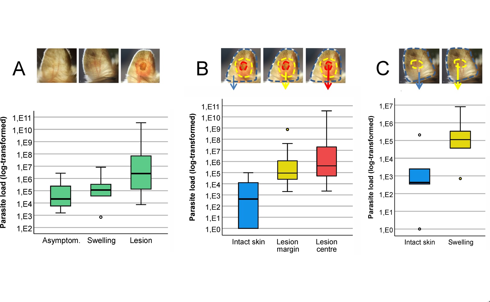
In its natural reservoir host, Shaw’s jird (Meriones shawi), Leishmania major parasites are heterogeneously distributed, concentrating in symptomatic areas of the ear (A) with parasite loads in lesion centers and margins being orders of magnitude higher than in the surrounding intact skin (B, C).

Image adapted from: Vojtková et al. / CC BY (Source)

Key Findings

  • In a study on leishmaniasis in North African rodents, parasites were clustered in skin lesions, with the edges being surprisingly more infectious to sand flies than the center
  • Only a tiny number of parasites (2-10) were needed to infect sand flies from these natural hosts, a dose 100 times lower than required in lab mice
Leishmaniasis is a group of neglected diseases caused by tiny parasites called Leishmania, which are transmitted to humans and animals through the bite of infected sand flies. These diseases can manifest in various ways, from skin sores (cutaneous leishmaniasis) to severe, potentially fatal internal organ infections (visceral leishmaniasis). A major challenge in controlling leishmaniasis is understanding how the parasites are transmitted from an infected host to a sand fly, specifically, what makes a host "infectious" to the biting insect. While it was once thought that visible skin lesions were the primary source of infection for sand flies, and that parasites circulating in the blood (parasitemia) were the main determinant of transmission for visceral leishmaniasis[2], recent research has begun to shift this understanding. It has become clear that many infected individuals, particularly those with visceral leishmaniasis, remain asymptomatic, meaning they show no outward signs of disease, yet can still harbor the parasite and potentially transmit it, making them significant, hidden reservoirs[3][4]. A recent study conducted by researchers at Charles University, U. Reims, and U. Paris-Saclay[1] aimed to shed more light on these critical factors, specifically for Leishmania major, the cause of cutaneous leishmaniasis. The team investigated how parasites are distributed within the host's body and the minimum number of parasites needed for a sand fly to become infected. They focused their efforts on two animal models: Meriones shawi, a natural North African rodent reservoir for L. major, and BALB/c mice, a common laboratory model. To understand the parasite distribution, the scientists used a combination of techniques. Quantitative Polymerase Chain Reaction (qPCR) was employed to measure the amount of Leishmania DNA in different parts of the infected ear skin, allowing them to quantify the parasite load. They also used fluorescence microscopy to visually locate and count the parasites. To assess how infectious different areas of the skin were, they utilized a method involving minimally invasive microbiopsies. These tiny skin samples were designed to mimic how a sand fly feeds, penetrating the skin to a shallow depth and absorbing skin cells and fluid, similar to what a sand fly would ingest[4]. This allowed them to analyze the parasite content at specific bite sites and correlate it with whether sand flies feeding on those areas became infected. The study's findings revealed a complex picture of parasite distribution and infectiousness. Consistent with observations for visceral leishmaniasis[2], the L. major parasites were not uniformly spread throughout the infected ear. Instead, their distribution was "heterogeneous," meaning they were clustered in patches, with noticeable differences in concentration between the center of a lesion, its edges (margins), and the surrounding skin that appeared visually healthy. The research confirmed that sand flies did not become infected if they fed on areas where no parasites were detected by microscopy. Crucially, in the natural host, Meriones shawi, the lesion margins emerged as the most effective source of infection for sand flies. This is a significant finding because it suggests that even areas around a visible sore, or skin that might not look heavily infected, can be highly transmissible. This aligns with earlier findings that asymptomatic animals can be infectious weeks before skin lesions appear and months after they heal, indicating that visible lesions are not a prerequisite for vector infection[3]. The study further quantified the minimum number of parasites, called amastigotes (the form of the parasite found within host cells), required to infect a sand fly. In M. shawi, as few as 2 to 10 amastigotes at a bite site were sufficient to initiate infection in a sand fly, with successful infections occurring at sites containing 4 to 500 amastigotes. This remarkably low infection threshold was further confirmed through experiments where sand flies were fed through a special membrane containing known parasite numbers. A striking difference was observed when comparing the natural host to the laboratory BALB/c mouse model. In BALB/c mice, there were only minor differences in infectiousness between the center and margins of lesions. Furthermore, the minimum infectious dose in BALB/c mice was approximately 100 times greater than in M. shawi, requiring between 1,500 and 10,000 amastigotes for successful sand fly infection. These findings significantly advance our understanding of Leishmania major transmission. They highlight that the "skin parasite landscape" – where parasites are located and how they are distributed – is a critical predictor of how infectious a host is[2]. The discovery of a very low infectious dose in natural hosts, coupled with the high infectiousness of lesion margins and visually unaffected skin, underscores the importance of asymptomatic carriers as a source of infection, reinforcing previous observations[3][4]. This detailed knowledge is vital for developing more accurate mathematical models of cutaneous leishmaniasis epidemiology, which can then inform public health strategies. Moreover, the stark differences observed between the natural host and the standard laboratory mouse model emphasize the critical need to incorporate natural host models in research. The dynamics of disease progression and transmission parameters can vary significantly between species, meaning that relying solely on laboratory models might lead to an incomplete or even misleading understanding of disease transmission in real-world settings. This deeper insight into host infectiousness, particularly the role of skin parasites, is crucial for assessing the effectiveness of treatments and the impact of leishmaniasis elimination campaigns[2].

MedicineEcologyAnimal Science

References

Main Study

1) Infectiousness of Leishmania major to Phlebotomus papatasi: differences between natural reservoir host Meriones shawi and laboratory model BALB/c mice

Published 16th June, 2025

https://doi.org/10.1371/journal.pntd.0013183


Related Studies

2) Skin parasite landscape determines host infectiousness in visceral leishmaniasis.

https://doi.org/10.1038/s41467-017-00103-8


3) Infectiousness of Asymptomatic Meriones shawi, Reservoir Host of Leishmania major.

https://doi.org/10.3390/pathogens12040614


4) Minimally invasive microbiopsies: a novel sampling method for identifying asymptomatic, potentially infectious carriers of Leishmania donovani.

https://doi.org/10.1016/j.ijpara.2017.02.005


Related Articles

An unhandled error has occurred. Reload 🗙