Anesthesia Isn’t Sleep: Brain Activity Under Isoflurane Differs From Sleep

Greg Howard
1st June, 2025

Anesthesia Isn’t Sleep: Brain Activity Under Isoflurane Differs From Sleep

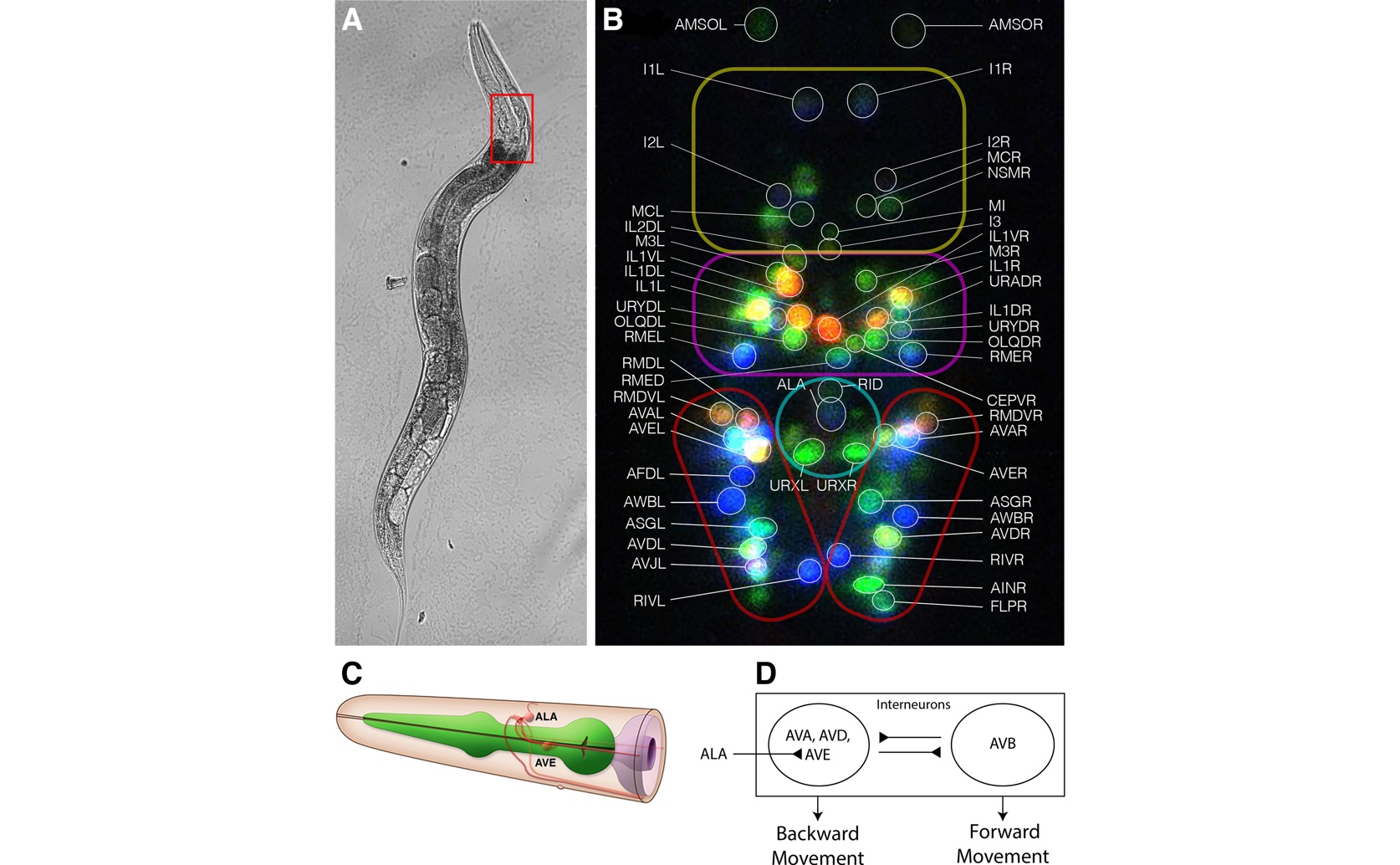
To establish the anatomical and functional baseline for testing whether anesthesia mimics sleep, this figure details the gross anatomy (A) and NeuroPAL-identified neurons (B) of Caenorhabditis elegans, highlighting the physiological circuit (C, D) where the ALA neuron inhibits the AVE interneuron to produce sleep-associated immobility—a mechanism the study ultimately demonstrates is distinct from that of isoflurane anesthesia.

Image adapted from: Chang et al. / CC BY (Source)

Key Findings

  • In C. elegans worms studied at Boston and Nebraska, natural sleep turns on neurons ALA and RIS and turns off the movement neuron (AVE) to keep the worm still
  • Under isoflurane anesthesia, this normal sleep pattern reverses—ALA and AVE activities become aligned, showing a different mechanism from natural sleep
[1] Researchers from Boston University School of Medicine, Brigham and Women’s Hospital, and the University of Nebraska Medical Center have recently investigated a fundamental question in neuroscience: do the same neuronal circuits underlie immobility in natural sleep-like states and in anesthesia? Using the nematode Caenorhabditis elegans—a model organism with a mapped nervous system—the study compared the activity patterns of key neurons during natural quiescence with those during isoflurane anesthesia. Sleep and anesthesia both result in immobility, but research has long suggested that the underlying brain mechanisms may be distinct. In C. elegans, behavioral quiescence, or sleep-like state, is characterized by a well-defined pattern of neuronal activation. In natural quiescence, specific neurons such as ALA, a GABAergic neuron, and another interneuron known as RIS become strongly active while other neurons, including command interneurons like AVE, are suppressed. The current study aimed to determine if the same circuit dynamics are involved in the immobility produced by anesthetic agents. To address this, the researchers used a combination of calcium imaging and a specialized neuronal labeling system. They employed a fluorescent calcium indicator called GCaMP6s, which glows when neurons are active, and captured neuronal activity using light-sheet microscopy. Neuronal identities were confirmed with the NeuroPAL nuclear labeling system. The study focused on the “sleep atonia pathway” of C. elegans, tracking activity in neurons such as ALA, AVE, and AVA as the animals were exposed to increasing levels of isoflurane anesthesia. Previous studies have demonstrated that the sleep-like state in C. elegans reproduces several hallmarks of sleep found in higher organisms, including behavioral quiescence, homeostasis (meaning the state is maintained by feedback mechanisms), and rapid reversibility[2]. Other research has highlighted common genetic elements regulating sleep across species, including a role for cGMP-dependent protein kinases in sensory neurons that promote sleep-like behavior in both nematodes and fruit flies[3]. Additionally, work investigating the role of interneurons in sleep regulation identified the RIS neuron as a critical component in promoting sleep through neuropeptide signaling, further underscoring the complexity of sleep circuitry in simple organisms[4]. In natural sleep-like quiescence, the study observed that neurons such as ALA and RIS display robust activation, while AVE, an important motor command interneuron, remains suppressed. Under these conditions, a moderate negative correlation exists between the activity of ALA and AVE (with a Pearson correlation coefficient around –0.286). This negative correlation is interpreted as a sign that when the sleep-promoting neuron is active, the command interneuron facilitating motion is inhibited, thereby promoting immobility. However, the pattern drastically changes under conditions of isoflurane anesthesia. As the depth of anesthesia increased, the researchers noted a progressive disruption of the typical negative correlation between ALA and AVE. Instead of maintaining the inhibitory relationship seen during natural quiescence, there was an inversion leading to a significantly positive correlation (r = 0.229, p = 0.003, as determined by ANOVA). In simpler terms, the activity between the sleep-promoting neuron and the command interneuron became more aligned rather than oppositional. Moreover, the normally strong positive correlation seen between AVE and another motor interneuron, AVA, was greatly diminished during deep anesthesia. These results indicate that while both sleep and anesthesia result in immobility, the underlying neuronal mechanisms differ. In natural sleep-like states, the coordinated activation of ALA and RIS along with the suppression of other neurons like AVE is crucial for establishing the state of immobility. In contrast, anesthesia does not mimic this natural pattern. The altered relationship between ALA and AVE under isoflurane suggests that the anesthetized state recruits an entirely different set of circuit dynamics to produce immobility. By revealing these differences, the study challenges the assumption that anesthetic-induced immobility uses the same neuronal pathways as natural sleep. It emphasizes that although the end result—lack of movement—is similar, the inner workings of the nervous system during these states are distinct. This finding is important because it cautions against oversimplifying the relationship between sleep and anesthesia and suggests that treatments or drugs targeting one state may not necessarily work the same way in the other. The new evidence ties together observations from earlier work in important ways. For instance, the natural sleep-like state observed in prior studies[2] and the genetic underpinnings of sleep-like behavior [3,4] are underscored by this study’s demonstration that immobility during sleep is a regulated phenomenon. Yet, when anesthesia is applied, the expected regulatory balance between sleep-promoting and motor command neurons breaks down. This divergence could help guide future research, particularly when exploring how anesthetics affect brain activity and how these effects differ from the natural sleep state. In summary, the study demonstrates that immobility in anesthetized C. elegans does not arise from the same neuronal mechanism seen in natural sleep-like quiescence. The nuanced differences in neuronal correlations have significant implications for understanding how various states of immobility are produced and controlled by the nervous system. These insights contribute to a broader understanding of how sleep and anesthesia differ at a circuit level, informing both basic neuroscience and the potential clinical applications of anesthetics.

MedicineAnimal Science

References

Main Study

1) Anesthesia isn’t sleep: The neuronal dynamics of immobility in isoflurane-anesthetized C. elegans differ from the activity patterns of previously established sleep-like quiescent states

Published 30th May, 2025

https://doi.org/10.1371/journal.pone.0324323


Related Studies

2) Multilevel modulation of a sensory motor circuit during C. elegans sleep and arousal.

https://doi.org/10.1016/j.cell.2013.11.036


3) Lethargus is a Caenorhabditis elegans sleep-like state.

https://doi.org/10.1038/nature06535


4) An AP2 transcription factor is required for a sleep-active neuron to induce sleep-like quiescence in C. elegans.

https://doi.org/10.1016/j.cub.2013.09.028


Related Articles

An unhandled error has occurred. Reload 🗙