Studying Protein Changes in Tissue Regrowth

Greg Howard
11th February, 2025

Studying Protein Changes in Tissue Regrowth

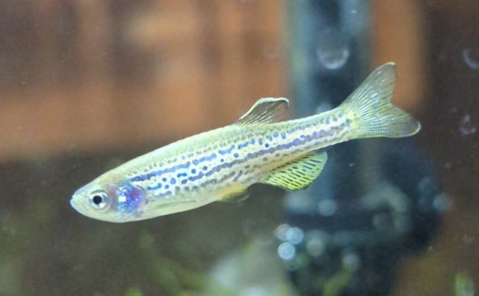
Zebrafish (Danio rerio)

Image Source: Wikimedia Commons

Key Findings

  • Researchers at CSIR-CCMB in Hyderabad studied how zebrafish regrow their fins by mapping protein changes
  • They found that specific proteins are active in the early days after fin injury, crucial for tissue regrowth
  • This discovery could help develop new treatments to promote tissue healing in humans
Regeneration, the process by which organisms repair or replace damaged tissues, has long intrigued scientists and philosophers. Zebrafish have emerged as a key model system for studying tissue regeneration due to their remarkable ability to regrow various body parts, including fins, brain, retina, spinal cord, and heart[2]. The genetic tools available for zebrafish research further enhance their suitability for uncovering the underlying mechanisms of regeneration. A recent study by researchers at CSIR-CCMB, Hyderabad, India[1] delves into the complex processes governing epimorphic regeneration in zebrafish caudal fins. Epimorphic regeneration involves the formation of a blastema, a mass of progenitor cells that proliferate and differentiate to reconstruct the lost tissue[3][4]. Understanding the molecular events within the blastema is crucial for deciphering how zebrafish achieve precise and efficient tissue regeneration. The research focused on elucidating protein phosphorylation patterns during fin regeneration. Protein phosphorylation is a post-translational modification where phosphate groups are added to proteins, altering their function and activity. By mapping these modifications, the study aimed to reveal the signaling pathways that drive the regeneration process. Using mass spectrometry analysis, the team identified a total of 440 phosphorylated proteins through immunoprecipitation with phosphoserine, phosphothreonine, and phosphotyrosine antibodies. Additionally, 74 phosphorylated proteins were detected using the TiO₂ column enrichment method, with 70 proteins showing differential phosphorylation during regeneration from 12 hours post-amputation (hpa) to 7 days post-amputation (dpa) compared to control samples. Remarkably, 95% of the proteins identified via the TiO₂ method were also found using the antibody pull-down approach, underscoring the reliability and accuracy of the findings. The researchers conducted whole mount immunohistochemistry to visualize phosphorylation events, revealing significant activity at 1 dpa, 2 dpa, and 3 dpa. This temporal pattern suggests that protein phosphorylation plays a critical role during the early stages of regeneration when the blastema is forming and expanding. Network pathway analysis highlighted that the differentially phosphorylated proteins are primarily associated with pathways related to cancer, organismal injuries, and abnormalities. This association indicates that the same signaling mechanisms involved in regeneration may also play roles in disease states, offering potential targets for therapeutic interventions. Previous studies have established that fin regeneration involves complex cellular interactions and signaling networks[2][3][4]. The current research builds on this foundation by providing a detailed map of phosphorylation events, thereby linking specific molecular modifications to the regenerative process. For instance, the identification of interleukin and solute carrier proteins from earlier transcriptomic analyses[5] complements the current findings by suggesting how these proteins might be regulated through phosphorylation during regeneration. The study's comprehensive approach, combining proteomics and advanced analytical techniques, offers a deeper understanding of the molecular choreography that enables zebrafish to regenerate their fins with precision. By revealing the specific proteins and pathways involved, this research paves the way for exploring how similar mechanisms might be activated or enhanced in mammals, potentially leading to breakthroughs in regenerative medicine. Moreover, the discovery of novel genes and proteins associated with regeneration provides new avenues for future research. Investigating these components could uncover additional layers of regulation and interaction that contribute to the regenerative capacity of zebrafish, further informing strategies to stimulate regeneration in other species[5]. In conclusion, the CSIR-CCMB, Hyderabad study significantly advances our knowledge of tissue regeneration by mapping phosphorylation patterns in zebrafish caudal fins. This work not only reinforces prior findings but also introduces new molecular targets that hold promise for developing regenerative therapies[2][3][4][5].

BiotechGeneticsBiochem

References

Main Study

1) Exploration of phosphoproteomic association during epimorphic regeneration.

Published 10th February, 2025

https://doi.org/10.1038/s41598-024-84735-z


Related Studies

2) The zebrafish as a model for complex tissue regeneration.

https://doi.org/10.1016/j.tig.2013.07.003


3) Old questions, new tools, and some answers to the mystery of fin regeneration.

Journal: Developmental dynamics : an official publication of the American Association of Anatomists, Issue: Vol 226, Issue 2, Feb 2003


4) The art of fin regeneration in zebrafish.

https://doi.org/10.1002/reg2.33


5) Understanding the complexity of epimorphic regeneration in zebrafish caudal fin tissue: A transcriptomic and proteomic approach.

https://doi.org/10.1016/j.ygeno.2022.110300


Related Articles

An unhandled error has occurred. Reload 🗙