New tool reveals detailed map of proteins inside cell power plants

Jenn Hoskins
19th December, 2025

New tool reveals detailed map of proteins inside cell power plants

Visualization of mitochondrial proteins with the BiG Mito-Split collection.

Image adapted from: Bykov et al. / CC BY (Source)

Key Findings

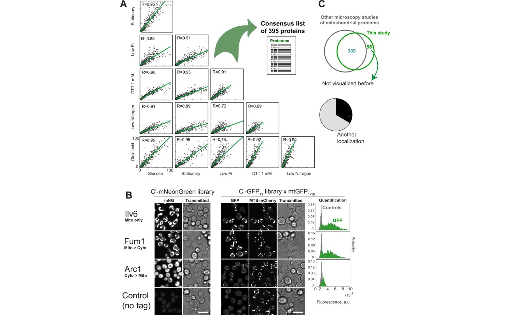
  • This study mapped approximately 400 mitochondrial proteins in yeast cells using a novel split-GFP assay, revealing 50 proteins not previously known to be in mitochondria
  • The assay identified dually localized proteins, meaning they reside in multiple cellular compartments, highlighting the complexity of protein distribution within cells
  • Researchers discovered an alternative start codon within the Gpp1 gene that generates a mitochondrial targeting signal, demonstrating hidden information can dictate protein localization
Mitochondria are often called the “powerhouses of the cell” because they generate most of the cell’s energy. However, these organelles are complex structures requiring many proteins to function, and understanding which proteins reside within them is a fundamental challenge in biology. Most mitochondrial proteins are actually made in the main body of the cell (the cytosol) and then imported into the mitochondria, not encoded directly within the mitochondria’s own DNA. Identifying all these proteins, particularly those without obvious “address labels” signaling their destination, has been difficult.[1] Researchers at the Weizmann Institute of Science recently tackled this problem, significantly expanding our knowledge of the mitochondrial protein landscape. The study focused on defining the complete “mitochondrial proteome” – the entire set of proteins found within mitochondria. A major hurdle has been identifying proteins that are dually localized, meaning they spend time in both the mitochondria and other parts of the cell. Traditional methods often struggle with these proteins because the signal from the mitochondria can mask the presence of the protein in other locations. To overcome this, the researchers built upon a technique they developed previously, called the Bi-Genomic split-GFP assay[2]. This assay cleverly splits the Green Fluorescent Protein (GFP) into two fragments. One fragment is encoded in the mitochondrial DNA, meaning it’s only produced inside the mitochondria. The other fragment is attached to the protein being studied. GFP only glows when the two fragments come together, ensuring that only proteins physically located inside the mitochondria are visible. To systematically identify mitochondrial proteins, the team used a strategy called SWAp-Tag (SWAT) to attach the GFP11 fragment (the second GFP fragment) to every protein in the yeast cell. They then combined these tagged proteins with the BiG GFP strain. By imaging the cells under different conditions, they were able to visualize almost 400 mitochondrial proteins, including 50 that had never been previously detected in mitochondria. Crucially, many of these newly identified proteins are dually localized, highlighting the complexity of protein distribution within the cell. This work builds on earlier advances in studying protein networks. The ability to easily manipulate and analyze proteins in simple organisms like yeast, combined with high-throughput cloning methods like the Gateway system[3], has been essential for these types of large-scale studies. The creation of comprehensive libraries of yeast strains with tagged proteins, as demonstrated in this study and others, provides valuable resources for the research community. The researchers didn’t stop at simply identifying the proteins. They investigated one of the dually localized proteins, Gpp1, in detail. Through structure-function analysis, they discovered an unexpected upstream start codon within the Gpp1 gene that generates a mitochondrial targeting signal. This finding illustrates how seemingly hidden information within a gene can dictate protein localization. Furthermore, they showcased how the BiG split-GFP assay can be used to study the complex process of how proteins are inserted into the inner mitochondrial membrane, a process where opposing mechanisms – stop-transfer and conservative sorting[4] – cooperate to ensure correct protein placement. The researchers made the library of GFP11-tagged strains freely available, which will be a powerful tool for future studies of protein localization, how proteins are made and transported within the cell (biogenesis), and how proteins interact with each other. This work represents a significant step towards completing the map of the mitochondrial proteome and understanding the intricate workings of these essential organelles.

GeneticsBiochemPlant Science

References

Main Study

1) A systematic bi-genomic split-GFP assay illuminates the mitochondrial matrix proteome and protein targeting routes

Published 16th December, 2025

https://doi.org/10.7554/eLife.98889


Related Studies

2) Assigning mitochondrial localization of dual localized proteins using a yeast Bi-Genomic Mitochondrial-Split-GFP.

https://doi.org/10.7554/eLife.56649


3) A suite of Gateway cloning vectors for high-throughput genetic analysis in Saccharomyces cerevisiae.

Journal: Yeast (Chichester, England), Issue: Vol 24, Issue 10, Oct 2007


4) Cooperation of stop-transfer and conservative sorting mechanisms in mitochondrial protein transport.

https://doi.org/10.1016/j.cub.2010.05.058


Related Articles

An unhandled error has occurred. Reload 🗙