Horse scar tissue composition reveals clues about healing speed

Greg Howard
8th November, 2025

Horse scar tissue composition reveals clues about healing speed

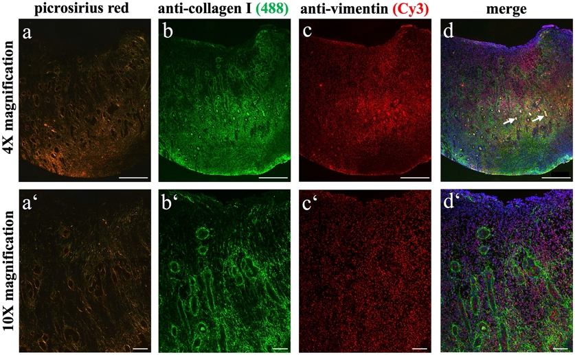
Collagen distribution, assessed by both Picrosirius red staining and immunofluorescence, reveals a higher density of mature collagen fibers in the deeper regions of equine exuberant granulation tissue wounds alongside evidence of vascular occlusion.

Image adapted from: Partusch et al. / CC BY (Source)

Key Findings

  • In horses, wounds especially on legs, often develop excessive tissue growth called exuberant granulation tissue (EGT), hindering healing and sometimes leading to euthanasia
  • EGT tissue contains significantly more immature type III collagen compared to normally healing tissue, suggesting a stalled healing process and impaired tissue maturation
  • Blood vessels within EGT are frequently blocked and cells lining them are enlarged, alongside changes in structural proteins, contributing to the abnormal tissue development
Wound healing in horses can be complicated, particularly when wounds develop excessive tissue growth known as exuberant granulation tissue (EGT). This condition, frequently seen on the lower legs, prolongs healing, increases treatment costs, and in severe cases, can lead to euthanasia[2]. While EGT is a common clinical problem, the underlying biological reasons for its development haven’t been fully understood, hindering the creation of effective treatments. Researchers from Ghent University, University of Nottingham, University of Veterinary Medicine Vienna, Leipzig University, and GH Raisoni College of Engineering and Management investigated the composition of EGT to better understand why it forms and how it differs from normal healing tissue[1]. The study focused on naturally occurring wounds in 19 horses with EGT, comparing them to six horses with normally healing wounds. Typically, when a wound heals, collagen – a protein providing structure – is laid down in a specific way. Initially, a more immature form of collagen, type III, is produced, which is later replaced by a stronger, more organized type I collagen. The research team examined the types of collagen present in both EGT and control wounds using a technique called histology, which involves examining tissue samples under a microscope. They found significantly higher levels of type III collagen in both the superficial (outer) and deep layers of EGT compared to the control wounds, suggesting that the healing process in EGT is stalled in an immature state. The total amount of collagen didn’t differ significantly between the groups, indicating the problem isn’t a lack of collagen, but rather the type of collagen being produced. Further investigation using immunofluorescence – a technique that uses antibodies to identify specific proteins – revealed additional differences. The researchers found that blood vessels within the EGT were often blocked and the cells lining them were enlarged. They also observed myofibroblasts, specialized cells involved in wound contraction and collagen production, were present throughout the EGT tissue. Importantly, the levels of certain proteins that provide structural support within cells were reduced in the superficial layer of EGT. These findings build upon earlier work highlighting the challenges of wound management in horses[3]. A survey of New Zealand veterinarians revealed that wounds, particularly those on the distal limb, are a significant clinical issue, often requiring prolonged and costly treatment, and sometimes leading to euthanasia. This study helps explain why these distal limb wounds are so problematic, pointing to a fundamental difference in the tissue composition during healing. The study also addresses a gap identified in a previous review of EGT treatments[4], which noted a lack of robust, evidence-based recommendations and a need for research using naturally occurring wounds in a larger number of horses. While the previous review highlighted the multifactorial nature of EGT, this new research provides specific insights into the collagen composition and vascular abnormalities contributing to the condition. The research suggests that promoting the maturation of the tissue – encouraging the switch from type III to type I collagen – could be a valuable therapeutic strategy. By understanding the specific differences in tissue composition, researchers can now focus on developing targeted treatments to improve wound healing and reduce the incidence of EGT in horses.

HealthBiochemAnimal Science

References

Main Study

1) Collagen composition in equine exuberant granulation tissue reflects tissue immaturity

Published 6th November, 2025

https://doi.org/10.1371/journal.pone.0335179


Related Studies

2) A cross-sectional survey on wounds in horses in New Zealand.

https://doi.org/10.1080/00480169.2015.1091396


3) BEVA primary care clinical guidelines: Wound management in the horse.

https://doi.org/10.1111/evj.13289


4) The granulation (t)issue: A narrative and scoping review of basic and clinical research of the equine distal limb exuberant wound healing disorder.

https://doi.org/10.1016/j.tvjl.2022.105790


Related Articles

An unhandled error has occurred. Reload 🗙