How Brain Cell Splicing Controls Nerve and Muscle Connections

Greg Howard
14th September, 2025

How Brain Cell Splicing Controls Nerve and Muscle Connections

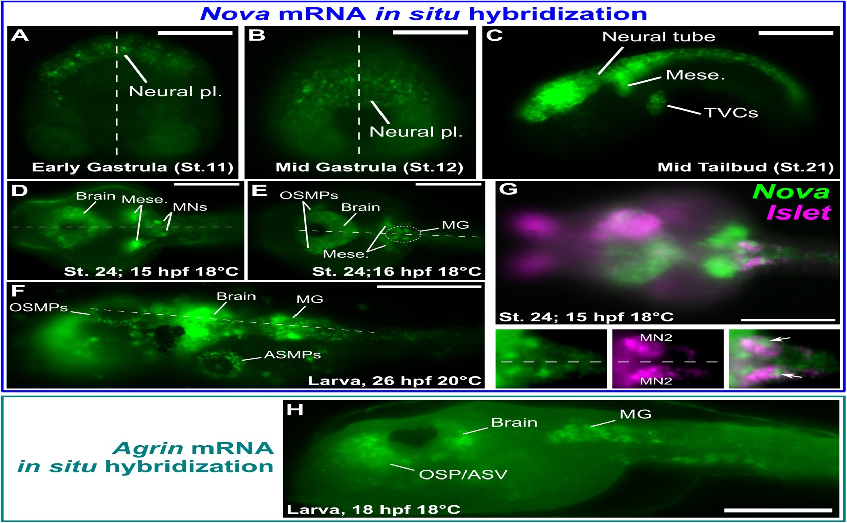
Fluorescent in situ hybridization demonstrates that Ciona robusta Nova is expressed in developing neural tissues (a–e), where it co-localizes with the motor neuron marker Islet (g) and spatially overlaps with Agrin transcription (h), establishing the cellular context for the proposed motor neuron-specific splicing pathway.

Image adapted from: Hossain et al. / CC BY (Source)

Key Findings

  • Nova protein, essential for nerve function, is expressed in motor neurons of Ciona robusta, a simple chordate
  • Nova protein promotes the inclusion of specific segments (Z exons) in Agrin mRNA, a process vital for nerve signal reception
  • The Nova-Agrin pathway for acetylcholine receptor clustering at nerve connections is conserved between Ciona and mammals
Amyotrophic lateral sclerosis (ALS) is a devastating neurological disease that progressively weakens muscles, eventually leading to paralysis and death. A key feature of ALS is the degeneration of motor neurons – the nerve cells that control muscle movement – and the connections they make with muscles at specialized junctions called neuromuscular junctions (NMJs)[2]. Understanding what goes wrong at the NMJ is crucial for developing effective treatments, as evidence suggests early problems here may trigger the entire disease process. Historically, it was thought that motor neuron degeneration initiated in the spinal cord and progressed outwards to the NMJ[2]. However, more recent research has challenged this view, with accumulating evidence indicating that the initial damage actually occurs at the NMJ itself, propagating “backwards” to the motor neuron cell body – a concept known as the “dying back” hypothesis[2][3]. This means that protecting or restoring NMJ function could potentially slow or halt the disease. The complexity of the NMJ, involving communication between the motor neuron, muscle, and supporting cells, makes it a challenging area to study, but also a promising therapeutic target. A new study conducted by researchers at St. John’s University, Georgia Institute of Technology, New York University, and University of Bergen[1] has shed light on a fundamental mechanism governing NMJ formation and maintenance, and importantly, demonstrates its surprising conservation across species. The research focuses on a protein called Agrin, which is essential for clustering acetylcholine receptors (AChRs) on the muscle surface. These receptors are vital for receiving signals from motor neurons, enabling muscle contraction. The process of making Agrin functional is complex. It begins with the pre-mRNA molecule for Agrin, which undergoes “alternative splicing”. This is a process where different parts of the mRNA are included or excluded during its conversion into a protein. The inclusion of small segments called “Z” exons is controlled by proteins called Nova1 and Nova2, which are specifically found in neurons. When Nova1/2 are present, they ensure the Z exons are included in the final Agrin protein, creating a neural-specific form that can bind to receptors (Lrp4) on the muscle cell and trigger AChR clustering. The study revealed that this entire pathway – alternative splicing of Agrin by Nova proteins to enable Lrp4-mediated AChR clustering – is remarkably similar in Ciona robusta, a simple marine animal that is a chordate (meaning it has a spinal cord) but is far less complex than mammals. To demonstrate this, the researchers used CRISPR/Cas9 gene editing in Ciona to remove the Nova gene. This led to a failure in Z exon inclusion and a loss of AChR clustering, confirming Nova’s essential role in this organism. They further showed that Ciona Nova protein could also function in mammalian cells to promote Z exon inclusion, proving its sufficiency. Interestingly, while the overall pathway is conserved, the researchers also found subtle differences in how Nova proteins function between Ciona and mammals. This suggests that the pathway has evolved over time, but the core mechanism remains the same. Furthermore, they identified a transcription factor called Ebf, which activates Nova expression in Ciona motor neurons. This links the RNA splicing process to a broader network of gene regulation specifically active in motor neurons. This study builds on earlier work showing pathological changes at the NMJ in ALS patients and mouse models[2][3]. The findings highlight the importance of the Agrin-Nova pathway in establishing and maintaining functional NMJs. The fact that this pathway is conserved in a simpler organism like Ciona makes it easier to study the underlying mechanisms and potentially identify new therapeutic targets. The research also suggests that disruptions in Nova expression or function could contribute to NMJ dysfunction in ALS, and that restoring Nova activity might be a viable strategy for treating the disease.

GeneticsBiochemEvolution

References

Main Study

1) Neuron-specific Agrin splicing by Nova RNA-binding proteins regulates conserved neuromuscular junction development in chordates

Published 12th September, 2025

https://doi.org/10.1371/journal.pbio.3003392


Related Studies

2) Neuromuscular Junction Dysfunction in Amyotrophic Lateral Sclerosis.

https://doi.org/10.1007/s12035-021-02658-6


3) Small junction, big problems: Neuromuscular junction pathology in mouse models of amyotrophic lateral sclerosis (ALS).

https://doi.org/10.1111/joa.13463


Related Articles

An unhandled error has occurred. Reload 🗙