Kaizen: Breaking Down Cell Images With an AI Technique

Jenn Hoskins
1st June, 2025

Kaizen: Breaking Down Cell Images With an AI Technique

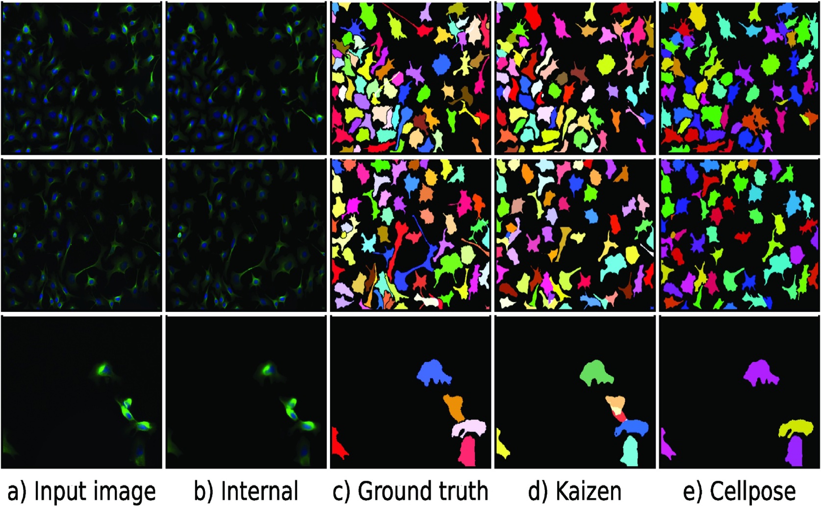
Qualitative evaluation on the neuroblastoma dataset demonstrates that Kaizen effectively decomposes original images (a) into accurate internal reconstructions (b) and segmentation masks (d), yielding object detection results comparable to ground truth annotations (c) and the Cellpose algorithm (e).

Image adapted from: Majoral et al. / CC BY (Source)

Key Findings

  • At the University of Tartu and CSIRO, researchers developed Kaizen, a new imaging method that predicts and refines cell boundaries in crowded microscope images
  • By iteratively updating an internal image model, Kaizen reduces errors and outperforms traditional methods in accurately identifying individual cells
A persistent challenge in cell and tissue biology is reliably finding and outlining cells in microscopy images, especially when cells are densely packed. Traditionally, methods rely on analyzing variations in pixel intensities to separate cells. However, these approaches often fail when boundaries between cells are not clearly defined due to tight clustering. In response to this difficulty, researchers at the University of Tartu and CSIRO developed an innovative approach called Kaizen[1]. The goal of Kaizen is to move beyond simple pixel-based segmentation, making object-level decisions that more effectively isolate individual cells in complex images. The Kaizen approach is built on the idea of maintaining an internal model of an image while comparing this model to the actual external image. This process draws inspiration from predictive coding—a theory in neuroscience suggesting that the brain continually predicts sensory input and revises those predictions based on discrepancies with incoming data. Kaizen replicates this principle computationally using a Vector Quantised-Variational AutoEncoder (VQ-VAE), a type of machine learning model that compresses and reconstructs image data by representing it with a limited set of predefined codes. In practice, Kaizen begins with an internal representation of the microscopy image. The system then iteratively examines the image, focusing on areas that do not match this internal model. For each area of discrepancy, the VQ-VAE makes a new “guess” about what the cell or structure might be. These candidate reconstructions are compared with the actual image data, and only those that bring the internal model closer to the external reality are retained. This iterative process continues until the internal model adequately reflects the input image, effectively decomposing the densely packed image into its constituent cells. Kaizen was tested on two fluorescence microscopy datasets. Fluorescence microscopy is a technique in which cells are stained with fluorescent dyes, causing them to emit light when excited by certain wavelengths. This enhances visibility of different structures in the cells—important when trying to distinguish components like nuclei and neuronal cells in dense cultures. The study demonstrated that Kaizen could improve the separation of these elements compared with traditional methods. By refocusing the segmentation process on entire cell objects rather than isolated pixels, Kaizen was able to make more consistent and accurate identifications in challenging images. It is worth noting that earlier work in this area, such as the development of Cellpose[2], also advanced the field of image segmentation. Cellpose introduced a deep learning method that could directly segment cells across a wide variety of image types without needing extensive retraining or parameter adjustments. Although Cellpose has been successful by training on vast and diverse datasets of images, its approach typically focuses on pixel-level intensity information. In contrast, Kaizen builds on and extends these ideas by integrating object-level decision making—a move that marks a notable evolution from simply using preset rules for pixel analysis. While Cellpose relies on having a large dataset upfront, Kaizen uses an iterative, predictive approach that dynamically adapts the internal representation of the image to ensure that object boundaries are correctly identified. A major strength of the Kaizen methodology lies in its adaptability. By continuously refining its internal model based on areas where predictions fall short, the system can handle the variability in cell shapes, sizes, and densities that is typical in biological samples. This flexibility is particularly important when dealing with fluorescence microscopy images that can range widely in quality and complexity, depending on the staining procedures and imaging conditions. Instead of depending solely on a static dataset or rigid segmentation rules, Kaizen’s iterative process allows it to self-correct during the segmentation task. This capability represents a significant step forward in the effort to develop universally applicable image segmentation methods for biological research. The work from the University of Tartu and CSIRO illustrates how drawing inspiration from neural processes can lead to practical computational strategies. Breaking away from traditional pixel-based analysis, Kaizen demonstrates the power of an approach that treats image segmentation as an evolving hypothesis-testing process. By incorporating ideas such as predictive coding from neuroscience, the method brings a new level of sophistication to cell segmentation tasks. This is especially valuable for research areas where accurate cell identification is critical, such as in studies of tissue organization, developmental biology, and cancer research. Researchers involved in the study believe that the Kaizen framework, with its internal representation and iterative improvement scheme, could be further refined and potentially adapted for other types of image analysis beyond biological microscopy. The iterative nature of the VQ-VAE used in Kaizen means that the method could improve over time as it processes more data and refines its internal model. This is a promising direction for future research and could lead to more robust tools across a range of scientific disciplines, enhancing our capability to analyze complex image data. Overall, the Kaizen approach marks a notable advance in the segmentation of microscopic images by integrating concepts from neuroscience and leveraging modern machine learning techniques. By focusing on object-level decisions and employing an iterative refinement process, it offers a promising alternative to traditional pixel-based segmentation methods. This new strategy may lead to more accurate and reliable analysis of densely packed cells, addressing long-standing challenges in cell and tissue biology.

Biotech

References

Main Study

1) Kaizen: Decomposing cellular images with VQ-VAE

Published 30th May, 2025

https://doi.org/10.1371/journal.pone.0313549


Related Studies

2) Cellpose: a generalist algorithm for cellular segmentation.

https://doi.org/10.1038/s41592-020-01018-x


Related Articles

An unhandled error has occurred. Reload 🗙