Protein Helper proSAAS is Abundant in the Eye's Retina

Jenn Hoskins
19th May, 2025

Protein Helper proSAAS is Abundant in the Eye's Retina

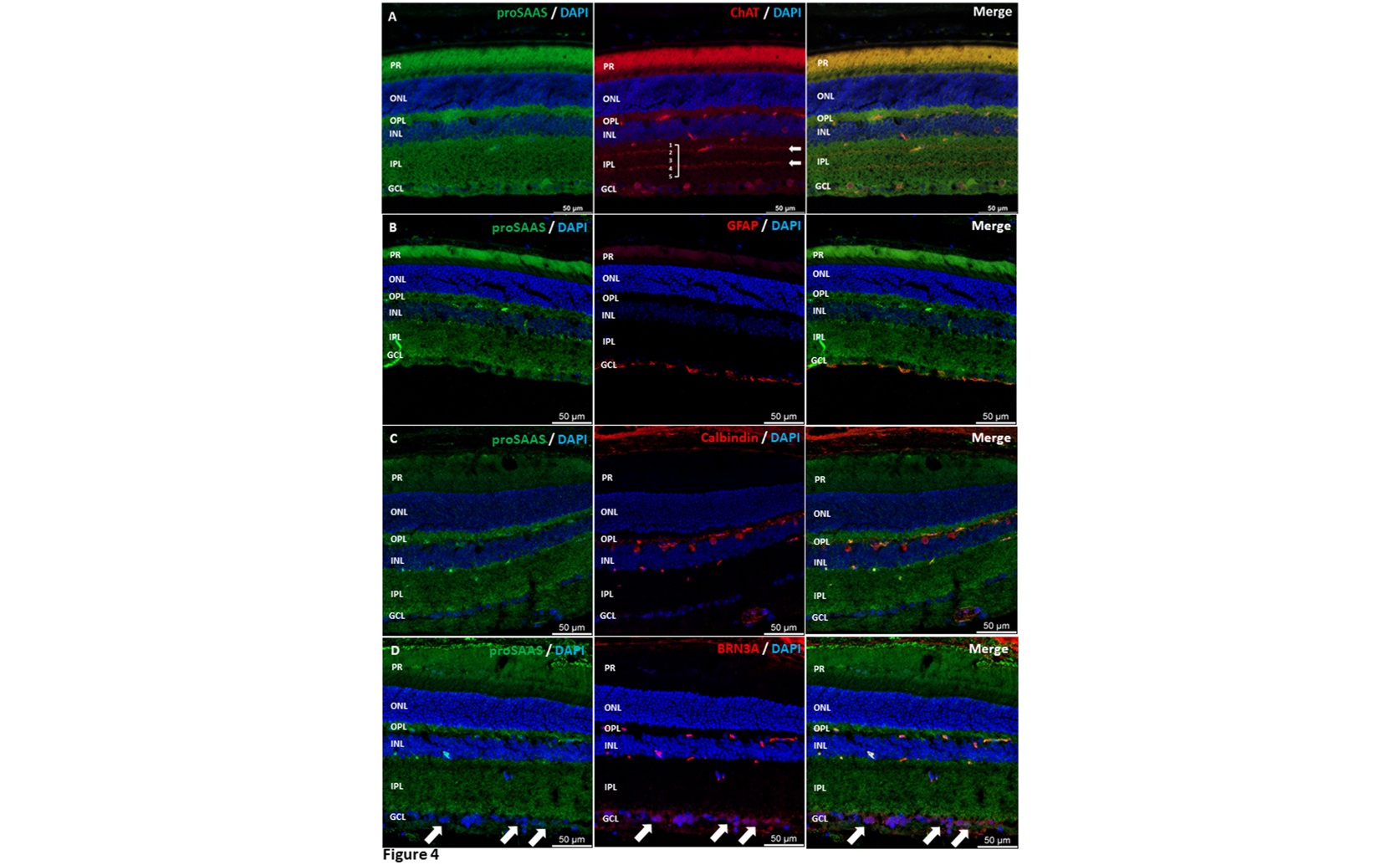
Confocal microscopy of mouse (Mus musculus) retinal sections demonstrates the localization of the chaperone proSAAS in amacrine cells (a) and astrocytes (b), with significant enrichment observed in horizontal cells (c) and retinal ganglion cells (d).

Image adapted from: Schaffer et al. / CC BY (Source)

Key Findings

  • *University of Maryland researchers found that the protein proSAAS is highly present in key cells of the retina, essential for maintaining clear vision.*
  • *ProSAAS helps other proteins fold correctly and prevents harmful clumps, ensuring the retina processes visual information effectively.*
  • *Boosting proSAAS could lead to new treatments for eye diseases like age-related macular degeneration and glaucoma.*
Maintaining clear vision is essential for daily life, but various diseases can impair this vital sense. The retina, a critical part of the eye, consists of specialized neurons that process visual information and send it to the brain. For the retina to function properly over a lifetime, it relies on a delicate balance of protein management, known as protein homeostasis or proteostasis[2][3]. Disruptions in proteostasis can lead to retinal diseases such as retinitis pigmentosa and age-related macular degeneration, which may result in vision loss or blindness. Recent research from the University of Maryland School of Medicine[1] has shed light on a specific protein, proSAAS, that plays a significant role in maintaining proteostasis within the retina. ProSAAS is a small protein known as a chaperone, which helps other proteins fold correctly and prevents them from clumping together. In the study, scientists discovered that proSAAS is highly expressed in certain retinal cells, particularly retinal ganglion cells (RGCs) and horizontal cells, which are crucial for processing and transmitting visual signals. Using advanced techniques such as RNA sequencing, the researchers analyzed both human and mouse retinas to determine where proSAAS is most active. They found that proSAAS is concentrated in the ganglion cell layer and the inner plexiform layer of the retina. These areas are dense with synapses, the connections between neurons that allow for the transmission of visual information. The presence of proSAAS in these layers suggests that it plays a key role in ensuring that the proteins involved in visual signaling remain functional and do not accumulate in harmful aggregates. Further analysis through Western blotting, a method used to detect specific proteins in a sample, revealed that proSAAS exists in two forms in the retina: a 21 kDa C-terminally processed form and a smaller 13 kDa fragment. The smaller fragment was also identified in human retinal samples, indicating that proSAAS undergoes processing to perform its functions effectively. This study builds on previous findings that highlight the importance of proteostasis in preventing retinal diseases. For example, disruptions in protein management have been linked to conditions like Stargardt's disease and glaucoma[2][4]. By identifying proSAAS as a key player in maintaining protein balance within retinal neurons, the research provides new insights into how the retina protects itself from protein-related damage. The methods used in this study were comprehensive and included both genetic and protein-level analyses. Single-cell sequencing allowed researchers to pinpoint which specific cells in the retina were producing proSAAS, while antibody techniques confirmed the protein's presence and distribution within retinal layers. These approaches provided a detailed picture of how proSAAS functions within the complex network of retinal neurons. Understanding the role of proSAAS opens up potential avenues for developing treatments for retinal diseases. If proSAAS helps prevent the aggregation of misfolded proteins, enhancing its activity or mimicking its function could be a strategy to protect retinal cells from degeneration. This aligns with previous research that has explored ways to bolster the proteostatic machinery to extend the functional lifespan of retinal cells[3]. In conclusion, the discovery of proSAAS's role in the retina adds a valuable piece to the puzzle of how proteostasis is maintained in this essential organ. By supporting the proper folding and functioning of proteins within retinal neurons, proSAAS helps safeguard against the development of degenerative eye diseases. Future research may focus on leveraging this knowledge to create therapies that strengthen proteostatic mechanisms, ultimately preserving vision and improving quality of life for those affected by retinal disorders.

MedicineHealthBiochem

References

Main Study

1) The neuronal chaperone proSAAS is highly expressed in the retina

Published 16th May, 2025

https://doi.org/10.1371/journal.pone.0321867


Related Studies

2) Protein misfolding and retinal degeneration.

https://doi.org/10.1101/cshperspect.a007492


3) Proteostasis in aging-associated ocular disease.

https://doi.org/10.1016/j.mam.2022.101157


4) Misfolded proteins and retinal dystrophies.

https://doi.org/10.1007/978-1-4419-1399-9_14


Related Articles

An unhandled error has occurred. Reload 🗙