Controlled Cancer Transformation in Single Cells Within Living Organisms

Greg Howard
28th March, 2025

Controlled Cancer Transformation in Single Cells Within Living Organisms

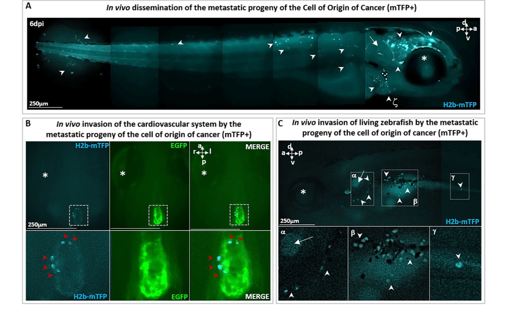
Confirming the malignant nature of the transformation, the progeny of a single cancerous brain cell in a zebrafish (Danio rerio) larva not only formed a primary tumor but also metastasized throughout the body (a), colonizing distant tissues like the heart (b) and digestive tract (c).

Image adapted from: Scerbo et al. / CC BY (Source)

Key Findings

  • Researchers in Paris used zebrafish to show that activating both a cancer-related gene and a cell-reprogramming factor in one cell can quickly form tumors
  • Turning on only the cancer gene KRASG12V didn’t cause cancer, highlighting that both factors are needed for a cell to become malignant
  • This controlled method helps scientists better understand how single cells turn cancerous, paving the way for improved cancer treatments
Cancer development often begins with normal cells acquiring genetic mutations in key genes that regulate cell growth and division. Understanding why some of these mutated cells transform into malignant tumors while others do not is crucial for developing effective cancer treatments. A recent study from Université de Paris, France[1] provides new insights into this process by demonstrating how specific genetic and cellular factors can drive a single cell to become cancerous. The research addressed the longstanding question of what triggers a normal cell with mutations in oncogenes or tumor suppressor genes to become malignant. Oncogenes are genes that, when mutated, can promote uncontrolled cell growth, while tumor suppressor genes normally help prevent cancer by regulating cell division and repairing DNA. The exact conditions under which these mutated cells turn cancerous have been difficult to pinpoint due to the unpredictable nature of malignant transformations. To overcome this challenge, the researchers employed an optogenetic approach in zebrafish, a model organism widely used in cancer research due to its genetic similarities to humans and transparent embryos that facilitate observation of cellular processes. They specifically activated an oncogene known as KRASG12V in a single cell within the zebrafish brain. KRASG12V is a well-known mutation associated with various cancers, including melanoma[2]. However, activating this oncogene alone was not sufficient to cause the cell to become cancerous. The breakthrough came when the researchers also transiently activated a reprogramming factor, VENTX/NANOG/OCT4, in the same cell. These factors are involved in maintaining pluripotency, a state where cells can develop into multiple types of cells during development[3]. The simultaneous activation of KRASG12V and the reprogramming factor led to a deterministic and reproducible transformation of the single cell into a malignant tumor within six days. This finding supports the "ground state theory of cancer initiation," which suggests that specific cellular states and interactions are critical for the initial steps of cancer development. This study builds on previous research that has explored the genetic and cellular contexts necessary for oncogenes to drive cancer. For instance, earlier work has shown that the SPRED1 gene, a negative regulator of MAPK signaling, is often inactivated in mucosal melanomas, leading to increased cell proliferation and drug resistance[2]. Additionally, studies on oncogenic competence have demonstrated that the ability of oncogenes like BRAFV600E to transform cells depends on the cell's intrinsic transcriptional programs and the presence of chromatin-modifying enzymes such as ATAD2[4]. These factors determine whether a cell is susceptible to transformation based on its developmental stage and genetic makeup. The integration of these insights into the main study highlights the importance of both genetic mutations and the cellular environment in cancer initiation. By manipulating both the oncogene and the reprogramming factor, the researchers were able to create a controlled scenario where a single cell reliably becomes cancerous. This approach not only clarifies the conditions necessary for malignant transformation but also provides a valuable model for studying cancer development and testing potential therapies. Furthermore, the study’s methodology of using zebrafish as a model organism allows for precise genetic manipulation and real-time observation of tumor formation. This technique complements previous findings on how specific genetic alterations and cellular states contribute to cancer, offering a platform to explore the interactions between different genetic factors and cellular environments[2][3][4]. In summary, the research from Université de Paris demonstrates that the combination of activating an oncogene and a reprogramming factor in a single cell can deterministically lead to tumor formation. This supports the idea that specific genetic and cellular contexts are essential for cancer initiation and provides a robust model for further investigating the mechanisms underlying malignant transformation.

MedicineHealthBiotech

References

Main Study

1) In vivo targeted and deterministic single-cell malignant transformation

Published 25th March, 2025

https://doi.org/10.7554/eLife.97650


Related Studies

2) Human tumor genomics and zebrafish modeling identify SPRED1 loss as a driver of mucosal melanoma.

https://doi.org/10.1126/science.aau6509


3) Vertebrate Cell Differentiation, Evolution, and Diseases: The Vertebrate-Specific Developmental Potential Guardians VENTX/NANOG and POU5/OCT4 Enter the Stage.

https://doi.org/10.3390/cells11152299


4) Developmental chromatin programs determine oncogenic competence in melanoma.

https://doi.org/10.1126/science.abc1048


Related Articles

An unhandled error has occurred. Reload 🗙