Gene and Protein Levels of Immune Markers in Uterine Cancer

Greg Howard
7th March, 2025

Gene and Protein Levels of Immune Markers in Uterine Cancer

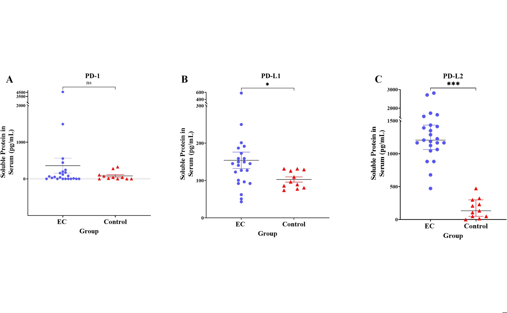
Serum analyses demonstrate that endometrial cancer patients have significantly elevated soluble PD-L1 (B) and PD-L2 (C), but not PD-1 (A), compared with controls, supporting the study’s conclusion that PD-L1 and PD-L2–mediated immune checkpoint activation is a prominent feature of endometrial cancer and a potential biomarker axis for immunotherapy stratification.

Image adapted from: Yusof et al. / CC BY (Source)

Key Findings

  • Researchers in Malaysia and Japan found that endometrial cancer tissues have much higher levels of PD-L1 and PD-L2 proteins compared to non-cancerous tissues
  • Elevated PD-L1 and PD-L2 are linked to more advanced cancer stages and lower survival rates in patients
  • These proteins may serve as important markers for predicting disease severity and as targets for personalized immunotherapy treatments
Endometrial cancer (EC), a prevalent malignancy of the female reproductive system, has traditionally been managed through surgical removal of tumors followed by chemotherapy, hormonal therapy, or radiation. Despite these treatments, outcomes often remain suboptimal, with high rates of recurrence and significant side effects. Recent advancements in personalized medicine have shifted the focus towards molecular analysis and innovative therapeutic strategies to improve patient outcomes[2]. A key area of interest in EC research is the role of immune checkpoints, specifically the programmed death-1 (PD-1) and programmed death ligands (PD-L1 and PD-L2) pathways. These molecules play a crucial role in regulating the immune system’s response to cancer cells. Normally, PD-1 on T cells interacts with PD-L1 or PD-L2 on other cells to prevent overactivation of the immune system. However, many cancer cells exploit this pathway to evade immune detection by increasing PD-L1 and PD-L2 expression, thereby inhibiting T cell activity and promoting tumor survival[3]. Building on this foundation, recent studies have explored the prevalence and impact of PD-L1 expression in EC. A systematic review revealed that approximately one-third of EC cases exhibit PD-L1 expression in tumor cells, and over half in immune cells, with higher expression linked to advanced disease stages and poorer survival outcomes[4][5]. These findings suggest that targeting the PD-1/PD-L1 axis could offer a promising therapeutic approach for EC patients, particularly those with advanced or aggressive disease forms. In this context, a recent study conducted by researchers at Universiti Kebangsaan Malaysia and Tohoku University[1] provides further insights into the molecular landscape of EC. This study delves into the expression levels of PD-1, PD-L1, and PD-L2 at both the mRNA and soluble protein levels in EC tissues compared to non-cancerous controls. By correlating these expression levels with various clinical profiles, the researchers aimed to identify potential biomarkers that could inform more effective immunotherapeutic strategies. The study found that EC tissues exhibited significantly higher mRNA expression and soluble protein levels of PD-L1 and PD-L2 compared to controls. Interestingly, PD-1 did not show a similar increase overall; however, its mRNA expression was notably higher in cases with less than 50% myometrial invasion, a measure of how deeply the cancer has penetrated the muscle layer of the uterus. Additionally, soluble PD-1 protein levels were significantly elevated in patients under 60 years of age. Further analysis revealed that higher PD-L1 gene expression was associated exclusively with advanced stages of EC. On the protein level, soluble PD-L1 was markedly increased in type II EC, which is typically more aggressive, as well as in cases with higher tumor grades, lympho-vascular space invasion (LVSI), and substantial myometrial invasion (50% or more). PD-L2 expression varied across most clinical profiles examined, except for LVSI. These findings expand upon previous research by not only confirming the elevated presence of PD-L1 in EC but also highlighting the nuanced roles of PD-1 and PD-L2 in different clinical contexts. The higher expression of PD-L1 and PD-L2 in more aggressive and advanced EC underscores their potential as biomarkers for disease prognosis and as targets for immunotherapy[2][4][5]. Specifically, the association of PD-L1 with poorer survival outcomes reinforces the rationale for using PD-L1 inhibitors in treating EC, aligning with earlier suggestions that immunomodulation could benefit patients with advanced disease[5]. Moreover, the differential expression of PD-1 in relation to myometrial invasion and age adds another layer of complexity, indicating that the immune environment in EC is influenced by multiple factors. This aligns with the broader understanding that immune checkpoint regulation is multifaceted, involving various mechanisms of protein expression and interaction[3]. By elucidating these patterns, the study provides a more detailed map of the immune landscape in EC, which is essential for developing targeted therapies. The methodologies employed in the study included quantitative analyses of mRNA and protein levels, which offer a comprehensive view of gene expression and protein availability in the tumor microenvironment. By integrating these molecular data with detailed clinical profiles, the researchers were able to identify specific associations that could inform personalized treatment approaches. This approach mirrors the personalized medicine paradigm discussed in earlier studies, where treatments are increasingly tailored based on individual molecular and genetic profiles[2]. In summary, the study by Universiti Kebangsaan Malaysia and Tohoku University advances our understanding of the immune mechanisms in endometrial cancer. By demonstrating the elevated levels of PD-L1 and PD-L2 and their association with more severe disease characteristics, the research supports the continued exploration of immune checkpoint inhibitors as a viable treatment option for EC. These findings contribute to the growing body of evidence that personalized immunotherapy, guided by specific biomarkers, holds significant promise for improving the prognosis and quality of life for patients battling endometrial cancer.

MedicineGeneticsBiochem

References

Main Study

1) Gene expression and soluble protein level of PD-1 and its ligands (PD-L1 and PD-L2) in endometrial cancer

Published 5th March, 2025

https://doi.org/10.1371/journal.pone.0312765


Related Studies

2) Expression of PD-1 and PD-L1 in Endometrial Cancer: Molecular and Clinical Significance.

https://doi.org/10.3390/ijms242015233


3) Immune checkpoint signaling and cancer immunotherapy.

https://doi.org/10.1038/s41422-020-0343-4


4) PD-L1 Expression in Endometrial Cancer and Its Association with Clinicopathological Features: A Systematic Review and Meta-Analysis.

https://doi.org/10.3390/cancers14163911


5) Programmed Death Ligand 1: A Poor Prognostic Marker in Endometrial Carcinoma.

https://doi.org/10.3390/diagnostics10060394


Related Articles

An unhandled error has occurred. Reload 🗙