How Salvia Root Extract Helps Prevent Heart Cell Death in Heart Failure

Greg Howard
3rd October, 2024

How Salvia Root Extract Helps Prevent Heart Cell Death in Heart Failure

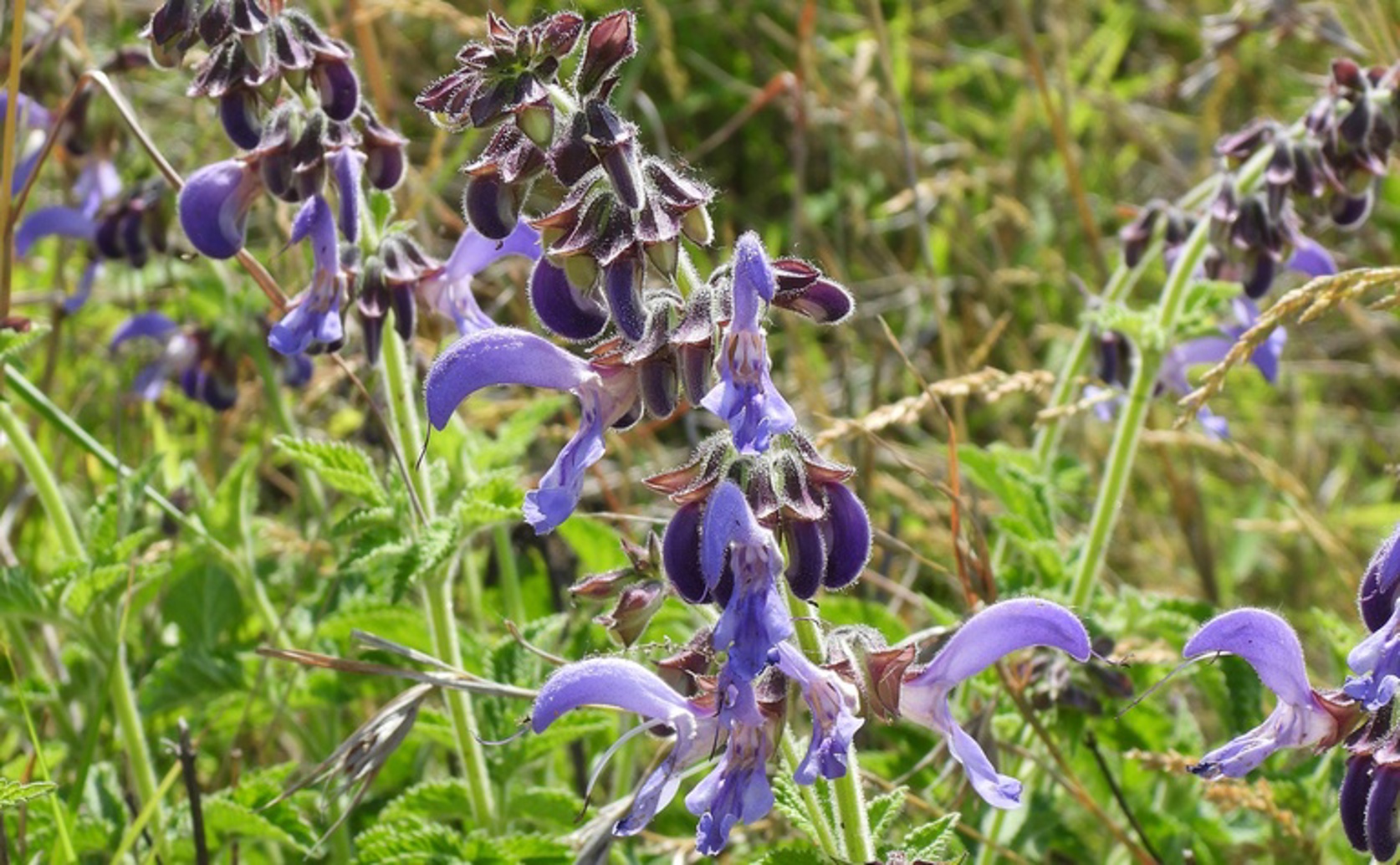
Salvia miltiorrhiza, source of Tanshinone IIA.

Photo adapted from: Chuangzao / CC BY-NC (Source). Used with permission.

Key Findings

  • The study from Changsha Fourth Hospital found that Tanshinone IIA (Tan IIA) can improve heart function in rats with heart failure
  • Tan IIA treatment reduced heart cell death and improved key heart function metrics like blood pressure and heart rate
  • The beneficial effects of Tan IIA are linked to restoring the PI3K/Akt/mTOR signaling pathway, which is crucial for cell survival and growth
Heart failure (HF) is a complex syndrome characterized by the heart's inability to pump sufficient blood to meet the body's needs. Acute heart failure (AHF) can manifest suddenly, either as a new condition or as a worsening of chronic heart failure, often leading to systemic congestion and organ dysfunction[2]. Despite advances in treatment, AHF remains associated with high mortality and hospital readmission rates due to the primarily symptomatic nature of current therapies[2]. Recent research from Changsha Fourth Hospital has investigated the potential of Tanshinone IIA (Tan IIA), a compound derived from Salvia miltiorrhiza Bunge (SMB), to improve cardiac function and reduce myocardial cell (MC) apoptosis in a rat model of heart failure[1]. The study involved fifty rats divided into five groups: a sham group (no treatment), a model group (HF model establishment), and three treatment groups receiving low, medium, and high doses of Tan IIA. The researchers assessed various indicators of cardiac function and MC apoptosis, as well as the levels of proteins associated with the PI3K/Akt/mTOR signaling pathway. This pathway is crucial for cell survival and growth, and its dysregulation has been linked to heart failure[3]. The results showed that the model group exhibited significant declines in several cardiac function metrics, including systolic arterial pressure, mean arterial pressure, heart rate, and left ventricular ejection fraction, compared to the sham group. Additionally, markers of MC apoptosis were significantly increased in the model group. However, the groups treated with Tan IIA demonstrated notable improvements in these indicators, suggesting that Tan IIA can enhance cardiac function and inhibit MC apoptosis. The PI3K/Akt/mTOR signaling pathway plays a pivotal role in mediating these effects. In the model group, the levels of phosphorylated PI3K, Akt, and mTOR proteins were significantly reduced, indicating impaired signaling. Treatment with Tan IIA restored the levels of these proteins, suggesting that Tan IIA exerts its protective effects by modulating this pathway. This study builds on earlier findings that highlight the significance of cellular mechanisms in heart failure. For instance, previous research has established that cardiomyocyte apoptosis, induced by factors like ischemia and hypoxia, contributes significantly to heart failure[4]. The role of integrin β3 in protecting against apoptosis and promoting cardiomyocyte proliferation through the PTEN/Akt/mTOR and ERK1/2 signaling pathways further underscores the importance of these molecular mechanisms in maintaining cardiac health[3]. In summary, the study from Changsha Fourth Hospital provides compelling evidence that Tan IIA can improve cardiac function and reduce myocardial cell apoptosis in heart failure by modulating the PI3K/Akt/mTOR signaling pathway. These findings not only offer a potential new therapeutic avenue for heart failure but also reinforce the critical role of molecular signaling pathways in the pathophysiology and treatment of this complex condition.

MedicineHealthBiochem

References

Main Study

1) Effects and mechanisms of Salvia miltiorrhiza Bunge extract on myocardial cell apoptosis in rat heart failure model.

Published 2nd October, 2024

https://doi.org/10.1590/acb396524


Related Studies


3) Integrin β3 promotes cardiomyocyte proliferation and attenuates hypoxia-induced apoptosis via regulating the PTEN/Akt/mTOR and ERK1/2 pathways.

https://doi.org/10.7150/ijbs.39414


4) Long Noncoding RNAs Involved in Cardiomyocyte Apoptosis Triggered by Different Stressors.

https://doi.org/10.1007/s12265-021-10186-w


Related Articles

An unhandled error has occurred. Reload 🗙