Cinnamon boosts bone cell growth in lab-made scaffolds for bone repair

Jim Crocker
3rd October, 2025

Cinnamon boosts bone cell growth in lab-made scaffolds for bone repair

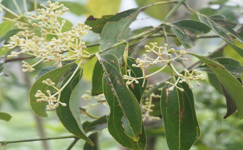
The delicate flowers of the Ceylon cinnamon tree (Cinnamomum verum), from which true cinnamon is harvested.

Photographer: Tero Linjama

Key Findings

  • Researchers in Zanjan, Iran, created fibers using PLLA and hydroxyapatite, adding either cinnamaldehyde or cinnamon essential oil to boost bone healing
  • Fibers with cinnamaldehyde showed the most promise, significantly increasing bone cell formation markers like calcium deposition and ALP activity
  • These fibers effectively balanced bone building and breakdown, suggesting a potential alternative to traditional bone grafts
Bone repair is a significant clinical challenge, particularly when defects are large or complex. Traditionally, the “gold standard” for repairing damaged bone involves taking bone from another part of the patient’s body – an autograft[2]. While effective, this procedure has drawbacks, including limited availability of donor bone and pain/complications at the donor site. Consequently, researchers are constantly seeking alternative materials and methods to stimulate the body’s natural bone regeneration processes. Researchers at Zanjan University of Medical Sciences recently investigated a novel approach using electrospun fibers to promote bone healing[1]. The study focused on creating a scaffold – a framework designed to support cell growth – made from poly-L-lactic acid (PLLA) and hydroxyapatite (HA), both biocompatible materials. Crucially, they incorporated either Cinnamaldehyde (Cin) or Cinnamon essential oil (E.O) into these fibers, aiming to create a dual-function system that both encourages bone formation and inhibits bone breakdown. The core issue the researchers addressed was achieving effective bone remodeling. Bone remodeling is a continuous process involving the coordinated action of cells that build new bone (osteoblasts) and cells that break down old bone (osteoclasts). A healthy balance is essential for maintaining bone strength and integrity. The researchers hypothesized that by incorporating Cin or E.O, they could modulate this process, promoting bone formation while simultaneously reducing excessive bone resorption. To create the scaffolds, the team used a technique called electrospinning. This process uses an electric field to draw charged threads of polymer solution, creating very fine fibers that mimic the natural extracellular matrix – the complex network surrounding cells in the body[3]. The resulting PLLA/HA fibers, whether loaded with Cin or E.O, were examined to ensure they had the desired characteristics: a smooth, bead-free structure suitable for cell attachment, and appropriate physical properties for supporting cell growth. The biocompatibility of the scaffolds was first tested using a standard cell viability assay (MTT assessment) to confirm that the materials weren’t toxic to cells. Following this, the team assessed the scaffolds’ ability to promote osteogenic – bone-forming – activity. This involved several tests, including measuring alkaline phosphatase (ALP) activity (an early marker of bone formation), quantifying calcium deposition (a key component of bone), and using a technique called Alizarin Red staining to visualize mineralized bone matrix. They also used RT-PCR analysis to investigate the expression of genes specifically involved in bone formation, such as ALP, Runx2, osteocalcin, and osteonectin. The results showed that both PLLA/HA/Cin and PLLA/HA/E.O fibers significantly enhanced osteogenic activity compared to a control group (scaffolds without Cin or E.O). Specifically, the PLLA/HA/Cin group demonstrated the most promising results, with significantly increased calcium deposition, higher ALP activity, and notably upregulated expression of osteogenic-related genes. The release of Cin from the fibers was also carefully monitored over 12 days, demonstrating a sustained release profile. These findings build upon prior research highlighting the potential of biomimetic scaffolds to restore tissue function[3]. While earlier studies have focused on simply providing a structural framework for bone cells, this study goes further by actively modulating the bone remodeling process. The incorporation of Cin and E.O demonstrates a strategy for enhancing the osteoinductive properties of bone substitutes[2], although the osteoinductivity observed is still less potent than that of bone morphogenetic proteins (BMPs)[2]. The dual-function approach, inhibiting resorption and promoting formation, is particularly noteworthy, as it mimics the natural balance observed during bone healing[4]. The use of mesoporous silicate nanoparticles (MSNs) to deliver therapeutic agents alongside a scaffold material, as demonstrated in[4], shares similarities with the approach taken in by incorporating Cin and E.O, both representing strategies to enhance the bioactive properties of bone tissue engineering scaffolds. The study concludes that PLLA/HA/Cin fibers represent a promising candidate for bone tissue engineering applications, offering a potential alternative to autografts and other conventional bone grafting materials.

MedicineBiotechPlant Science

References

Main Study

1) Cinnamaldehyde/cinnamon essential oil loaded poly-L-lactic acid/ hydroxyapatite fibrous scaffolds as osteogenic differentiation enhancing system for bone tissue engineering applications

Published 30th September, 2025

https://doi.org/10.1186/s13036-025-00547-3


Related Studies

2) Osteoinduction of bone grafting materials for bone repair and regeneration.

https://doi.org/10.1016/j.bone.2015.07.007


3) Biomimetic electrospun nanofibrous structures for tissue engineering.

Journal: Materials today (Kidlington, England), Issue: Vol 16, Issue 6, Jun 2013


4) Bone remodeling-inspired dual delivery electrospun nanofibers for promoting bone regeneration.

https://doi.org/10.1039/c8nr07329e


Related Articles

An unhandled error has occurred. Reload 🗙