Sappanwood's Role in Making Cancer-Fighting Copper Particles

Greg Howard
27th June, 2025

Sappanwood's Role in Making Cancer-Fighting Copper Particles

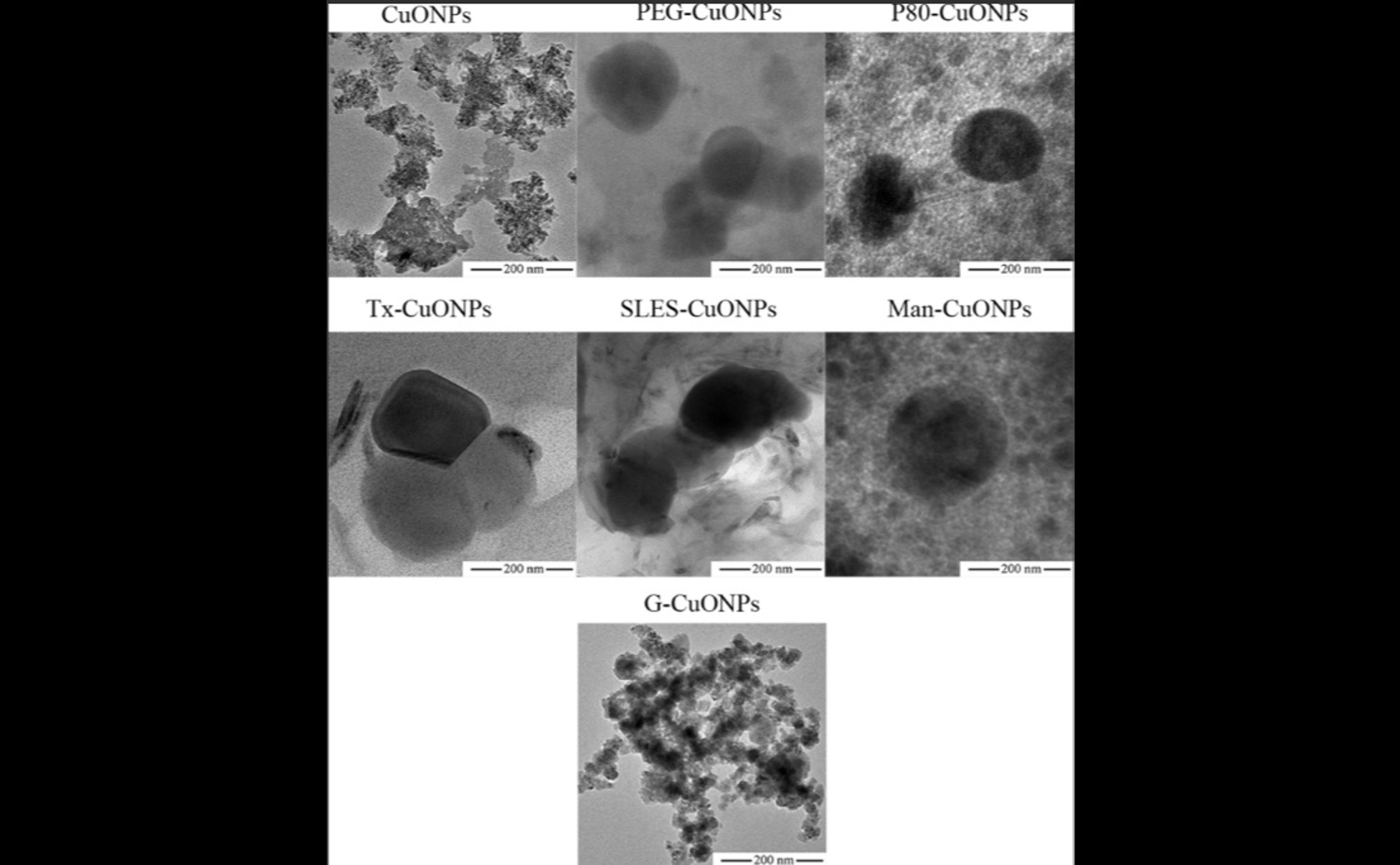
Transmission electron microscopy reveals that incorporating capping agents during biosynthesis with Caesalpinia sappan extract significantly reduces the strong agglomeration observed in uncapped copper oxide nanoparticles, ensuring the dispersed morphology and colloidal stability required for the study's demonstrated leukemic cytotoxicity.

Image adapted from: Sasarom et al. / CC BY (Source)

Key Findings

  • A study by researchers from Chiang Mai, Utrecht, and Al-Azhar Universities found that specific copper oxide nanoparticles can selectively kill leukemia cells
  • Specifically, copper oxide nanoparticles coated with PEG or P80 were highly effective at killing leukemia cells while sparing healthy cells, a key goal for safer cancer treatments
  • This selective killing ability was comparable to a standard chemotherapy drug, doxorubicin, offering a promising new direction for more targeted leukemia therapy
Cancer treatments have made significant strides, but many conventional approaches still face challenges, such as severe side effects and a lack of precise targeting. This often leads to damage to healthy tissues alongside cancerous ones. The field of nanomedicine offers a promising alternative, aiming to overcome these inherent shortcomings by delivering drugs more specifically to diseased cells, potentially reducing harm to the rest of the body[2]. However, despite the significant interest and attractive attributes of nanotechnologies, their journey from laboratory discovery to widespread clinical use, known as clinical translation, remains challenging. Experts have pointed out several critical hurdles that prevent or delay the adoption of nanomedicines. These include a limited understanding of how these tiny particles behave physically and chemically, difficulties in ensuring they reach only the intended cells or tissues, and a frequent inability to reproduce promising preclinical results in human clinical trials. Concerns about how well these materials interact with the body, known as biocompatibility, also persist[3]. Furthermore, the practical aspects of developing nanomedicines, such as scaling up production for industrial use, adhering to strict manufacturing standards, securing funding, and navigating complex regulatory pathways, add layers of complexity to their translation[3][4]. To accelerate success, it's crucial to consider the perspective of the end-stage users – patients and clinicians – early in the development process, ensuring that practical and clinical feasibility are central to design[4]. Addressing these challenges, a recent study by researchers from Chiang Mai University, Utrecht University, and Al-Azhar University[1] investigated a new approach to developing more effective and selective nanomedicines for leukemia, a type of blood cancer. The study focused on copper oxide nanoparticles (CuONPs), which are tiny particles made of copper and oxygen, typically measuring less than 100 nanometers, though in this study, the particles ranged from 175–280 nanometers. These nanoparticles were created using a natural extract from the Caesalpinia sappan plant, which acts as a "reducing agent" to help form the nanoparticles in a more environmentally friendly way, often referred to as biosynthesis. A key aspect of this research involved the use of "capping agents." These are substances added during the nanoparticle creation process that coat the surface of the nanoparticles. Capping agents are crucial because they help stabilize the nanoparticles, preventing them from clumping together, and can influence how they interact with biological systems in the body. For this study, several different capping agents were tested, including gelatin, polyethylene glycol 400 (PEG), and polysorbate 80 (P80), among others. The control group of CuONPs was prepared using gelatin as a capping agent and a different chemical reducing agent, sodium borohydride. To understand how these different capping agents affected the nanoparticles, the researchers first meticulously characterized their "physicochemical properties." This involved measuring their size and size distribution using dynamic light scattering, determining their surface electrical charge (known as "zeta potential"), and analyzing their chemical composition and structure using techniques like energy dispersive X-ray spectroscopy and Fourier-transform infrared spectroscopy. Understanding these properties is fundamental, as highlighted by previous research, to overcoming barriers in clinical translation[3]. The study found that most of the synthesized CuONPs had a size range of 175–280 nanometers, with a good size distribution, and a negative zeta potential (between -30 to -35 mV), which generally indicates good stability in a solution. The gelatin-capped particles, however, had a much less negative zeta potential of -3 mV, suggesting they might be less stable. The core of the study involved testing the "cytotoxic effects" of these nanoparticles. Cytotoxicity refers to the ability of a substance to kill cells. The researchers incubated the CuONPs with two types of cells: normal human peripheral blood mononuclear cells (PBMC), which serve as healthy control cells, and three different strains of leukemic cancer cells (KG1a, K562, and Molt4). The goal was to determine the "IC50 values," which represent the concentration of the nanoparticles required to inhibit the growth of 50% of the cells. A lower IC50 value indicates greater potency. The results were particularly promising for CuONPs capped with PEG and P80. These formulations demonstrated a significantly higher "selectivity index." The selectivity index is a crucial measure, calculated by dividing the IC50 value for healthy cells by the IC50 value for cancer cells. A higher selectivity index means the substance is more toxic to cancer cells than to healthy cells, which is a key objective in cancer therapy to minimize side effects. For PEG-CuONPs, the IC50 for healthy PBMC was 72.5 ± 5.8 µg/mL, while for leukemic cells, it ranged from 26–29 µg/mL. Similarly, for P80-CuONPs, the IC50 for healthy PBMC was 85.0 ± 3.1 µg/mL, and for leukemic cells, it was 28–41 µg/mL. These figures clearly show that both PEG- and P80-capped CuONPs were more effective at killing leukemic cells than healthy cells. This study directly contributes to fulfilling the "great promise" of nanomedicine for cancer treatment[2]. By identifying specific capping agents that enhance the selectivity of copper oxide nanoparticles against leukemia cells, the researchers are addressing critical aspects of nanomedicine development. The careful characterization of physicochemical properties helps to overcome the "poor understanding" barrier previously identified[3]. Furthermore, the focus on a high selectivity index is vital for improving "biocompatibility concerns" and designing effective "therapeutic endpoints" from an "end-user's perspective"[3][4]. The findings mark a significant step in the "preclinical development" phase, providing strong candidates for further investigation in living organisms. While challenges like industrial scale-up, regulatory approval, and funding still need to be navigated for widespread clinical success[3], this research offers a compelling direction for the development of more targeted and less harmful treatments for leukemia.

HerbsMedicineBiotech

References

Main Study

1) Influence of capping agents on physicochemical properties and leukemic cytotoxicity of copper oxide nanoparticles biosynthesized using Caesalpinia sappan extract

Published 26th June, 2025

https://doi.org/10.1371/journal.pone.0326791


Related Studies


3) A translational framework to DELIVER nanomedicines to the clinic.

https://doi.org/10.1038/s41565-024-01754-7


4) Challenges in nanomedicine clinical translation.

https://doi.org/10.1007/s13346-020-00740-5


Related Articles

An unhandled error has occurred. Reload 🗙