How A Gut Bacteria Toxin Forms Step-By-Step

Jim Crocker
22nd July, 2025

How A Gut Bacteria Toxin Forms Step-By-Step

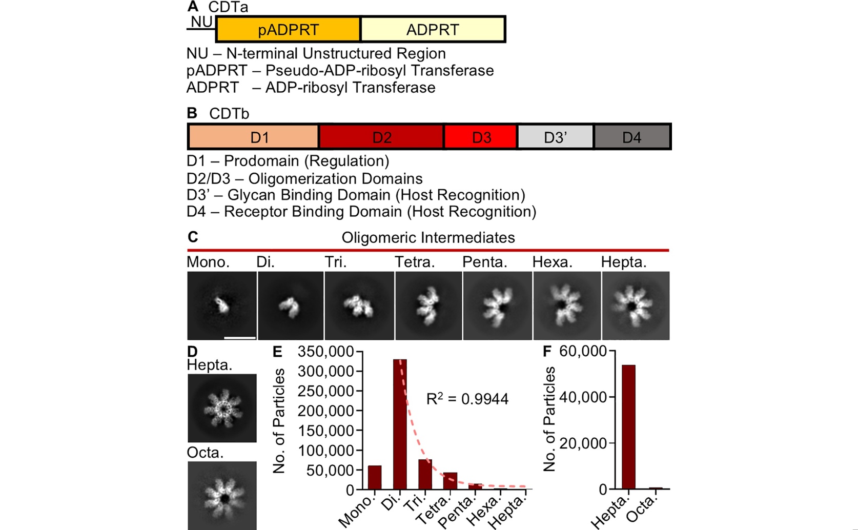
Cryo-EM analysis of the Clostridioides difficile transferase components (a, b) reveals various structural intermediates (c, d) whose particle distributions (e, f) demonstrate that toxin oligomerization proceeds through a stepwise mechanism.

Image adapted from: Mullard et al. / CC BY (Source)

Key Findings

  • Scientists at the University of Minnesota and University of Pittsburgh discovered how the C. difficile toxin (CDT) assembles step-by-step to cause severe illness
  • They found that the toxin's components change shape during assembly, a process triggered by water-repelling molecules and stabilized by another part of the toxin
  • This detailed understanding of toxin assembly provides crucial insights for developing new drugs to block CDT and combat severe C. difficile infections
Clostridioides difficile, often referred to as C. difficile, is a bacterium that has become the leading cause of infectious diarrhea acquired in hospitals across the United States. Infection with C. difficile can be severe and, in many cases, fatal. The bacterium produces and secretes up to three toxins: Toxin A, Toxin B, and a third, increasingly common toxin known as C. difficile transferase (CDT). While Toxin A and Toxin B are well-known drivers of the disease's pathology, strains of C. difficile that produce CDT have been consistently linked to more severe illness, higher rates of infection returning after treatment, and an increased risk of death. Earlier research has strongly supported the notion that CDT contributes significantly to the severity of C. difficile infection. For instance, a study comparing patients infected with C. difficile strains possessing genes for toxins A and B with those also possessing the gene for binary toxin (CDT) found that patients with CDT-producing strains had significantly higher case-fatality rates[2]. This was true regardless of the specific genetic type (PCR ribotype 027 or non-027) of the C. difficile strain, suggesting that CDT itself is either a marker for more virulent strains or directly enhances their harmfulness[2]. Further reinforcing this, another study demonstrated that the direct detection of CDT from patient stool samples correlated with more severe disease, a higher likelihood of complications, and increased recurrence rates[3]. When both CDT and Toxin B were detected simultaneously, their impact on prognosis was even greater[3]. The mechanism by which CDT enhances virulence has also been explored. Research indicates that CDT induces a harmful inflammatory response in the host, specifically through a pathway involving Toll-like receptor 2 (TLR2)[4]. This inflammatory response then suppresses a protective immune response normally provided by eosinophils, a type of white blood cell[4]. Restoring these protective eosinophils, even if they are TLR2-deficient, was shown to be sufficient for protection against CDT-producing strains in mice, offering a clear explanation for CDT's enhanced virulence and how it subverts the host's immune system[4]. Despite this growing understanding of CDT's clinical impact and its interaction with the immune system, a basic question remained: how does CDT itself assemble at a molecular level to intoxicate host cells? While it was known that the components of CDT must come together, or "oligomerize," to cause cellular damage, the precise molecular steps of this assembly process were not fully understood. Filling this crucial knowledge gap, recent research conducted by scientists at the University of Minnesota Twin Cities and University of Pittsburgh has provided unprecedented insights into the molecular assembly of CDT[1]. The researchers utilized a cutting-edge technique called cryogenic electron microscopy (Cryo-EM). Cryo-EM is a powerful imaging method that allows scientists to visualize biological molecules, such as proteins, at an incredibly detailed, near-atomic level. It works by flash-freezing samples, preserving their natural structure, and then using an electron beam to capture thousands of images, which are then computationally combined to create high-resolution 3D models. Using Cryo-EM, the team collected data from purified, recombinant CDT. From this data, they were able to generate several structural "snapshots" of the toxin, including a series of images that captured intermediate stages during its oligomerization. Oligomerization is the process where individual protein units come together to form a larger, functional complex. In the case of CDT, its two main components, CDTa and CDTb, must assemble to become an active toxin. These structural snapshots provided critical insight into the mechanism underlying the toxin's assembly, revealing a significant role for "structural plasticity" during this process. Structural plasticity refers to the ability of a molecule to change its shape. This flexibility is essential for the different parts of the toxin to fit together correctly and progress through the various stages of assembly. The study also demonstrated that even partially assembled toxins were equally potent in causing cell damage in laboratory tests, supporting their model of assembly in a cellular context. Furthermore, the researchers discovered that the larger component, CDTb, forms its oligomers more stably when CDTa is present, and that the assembly process is triggered by hydrophobic molecules. Hydrophobic molecules are those that repel water, and their involvement suggests specific chemical interactions drive the toxin's formation. This new understanding of how CDT assembles at a molecular level is profoundly significant. While previous studies established the clinical severity of CDT-producing C. difficile infections[2][3] and elucidated how the toxin subverts the host immune response[4], the precise molecular blueprint of the toxin's activation was missing. The work by the University of Minnesota Twin Cities and University of Pittsburgh provides this fundamental molecular description. By understanding the intricate steps of CDT assembly, including the role of structural plasticity and specific triggers like hydrophobic molecules, scientists can now identify potential weak points in the toxin's formation or function. This detailed molecular knowledge is crucial for developing new strategies to combat C. difficile infections, such as designing drugs that specifically block CDT's assembly or inhibit its activity, thereby mitigating the severe disease and high mortality rates associated with these highly virulent strains.

MedicineBiotechBiochem

References

Main Study

1) Oligomerization of the Clostridioides difficile transferase B component proceeds through a stepwise mechanism

Published 21st July, 2025

https://doi.org/10.1371/journal.ppat.1013186


Related Studies

2) Binary toxin and death after Clostridium difficile infection.

https://doi.org/10.3201/eid/1706.101483


3) The prognostic value of toxin B and binary toxin in Clostridioides difficile infection.

https://doi.org/10.1080/19490976.2021.1884516


4) The binary toxin CDT enhances Clostridium difficile virulence by suppressing protective colonic eosinophilia.

https://doi.org/10.1038/nmicrobiol.2016.108


Related Articles

An unhandled error has occurred. Reload 🗙