How Changes in a Protein Help Early Cell Development in Sea Urchin Embryos

Greg Howard
24th December, 2024

How Changes in a Protein Help Early Cell Development in Sea Urchin Embryos

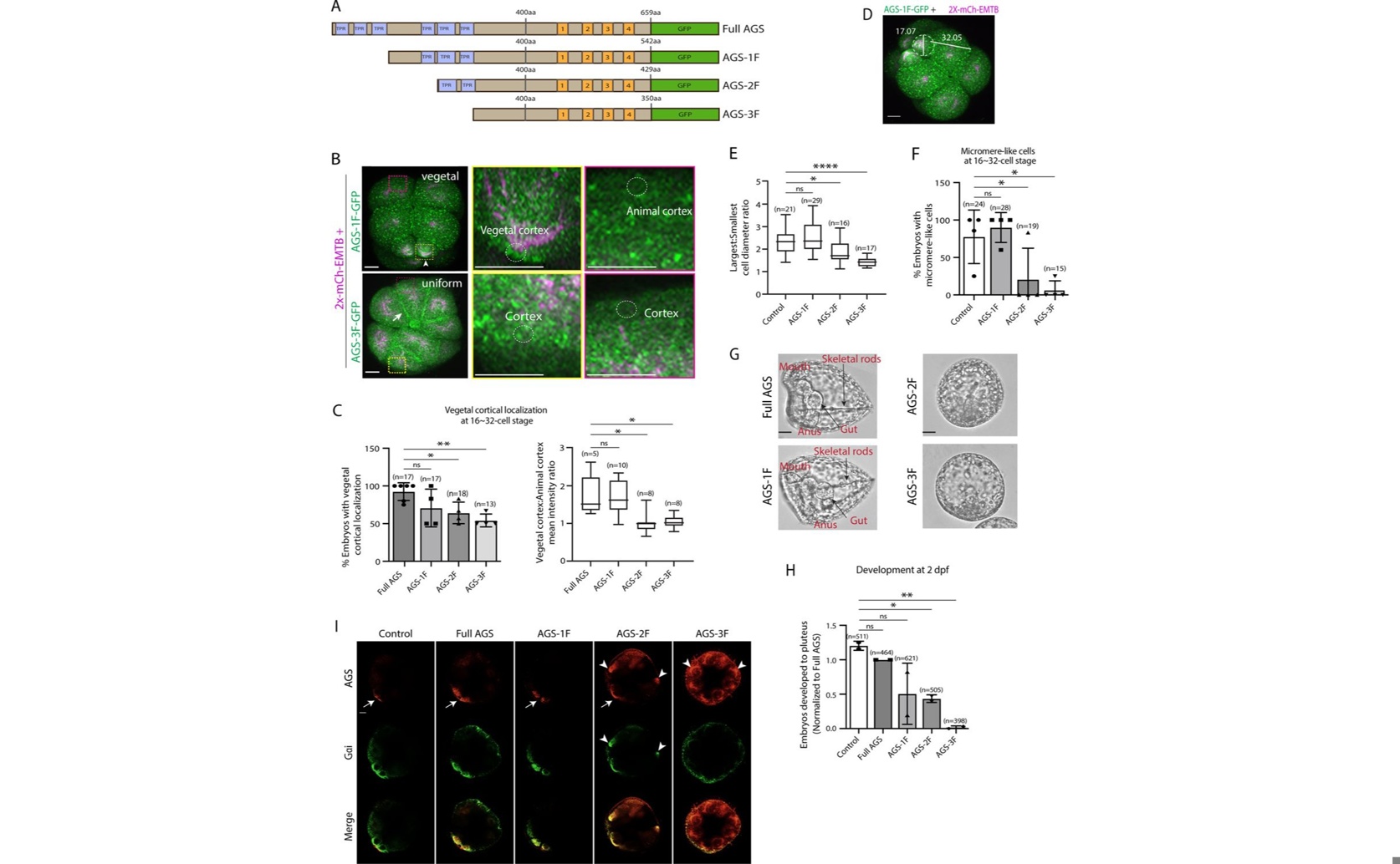
Deletion of the N-terminal TPR domain of the SpAGS protein in the sea urchin (Strongylocentrotus purpuratus) disrupts its crucial restriction to the vegetal cortex, causing uniform mislocalization (b, c, i) that prevents proper micromere formation (e, f) and subsequent embryonic development (h).

Image adapted from: Emura et al. / CC BY (Source)

Key Findings

  • Researchers at Brown University studied how sea urchin embryos develop unique cells called micromeres
  • They found that a protein called AGS is crucial for forming these micromeres during the 16-cell stage
  • The study showed that AGS has evolved uniquely in sea urchins, helping to create developmental diversity among echinoderms
The study conducted by researchers at Brown University delves into the evolutionary introduction of asymmetric cell division (ACD) in sea urchin embryos and how it contributes to developmental diversity and species diversification[1]. This research is pivotal as it explores the molecular mechanisms behind the formation of micromeres, a unique feature in sea urchins, and how these mechanisms have evolved. In sea urchin embryos, the 16-cell stage division results in the formation of micromeres, which are crucial for mesendoderm cell fate specification. This process is facilitated by a polarity factor known as AGS (Activator of G-protein Signaling). Interestingly, AGS and its associated ACD factors are present across various echinoderms and other metazoans, raising questions about what specific evolutionary modifications have led to the unique presence of micromeres in sea urchins. The study found that the GoLoco motifs at the C-terminus of the AGS protein play a significant role in regulating micromere formation in sea urchin embryos. The researchers examined AGS orthologs from other echinoderms, such as sea stars and pencil urchins, and observed that these orthologs showed varied localization and function in micromere formation. This suggests that while the core ACD machinery is preserved across echinoderms, the AGS protein has evolved uniquely in sea urchins to facilitate the formation of micromeres. Further, the study demonstrated that in the early embryogenesis of the pencil urchin, an ancestral type of sea urchin, the sea urchin AGS could facilitate micromere-like cell formation and accelerate the enrichment of the germline factor Vasa. This indicates that the molecular evolution of AGS has been a driving force in the diversity of ACD among echinoderms. The findings from this study build on previous research that has explored cell size asymmetries and cell fate decisions in echinoderms. For instance, earlier studies have shown that cell size asymmetries are consistently produced during sea star early cleavage and are predictive of the dorsoventral (DV) axis, although they are not necessary to instruct DV axis formation[2]. This highlights the complexity and variability in developmental processes among echinoderms. Additionally, research on the evolution of developmental gene regulatory networks (GRNs) in sea stars and sea urchins has shown that transcription factors like Tbrain have evolved significantly different roles in these species, with extensive evolutionary changes in their binding motifs and target genes[3]. This aligns with the current study's findings on the evolutionary modifications of AGS and its impact on ACD diversity. In conclusion, the study from Brown University provides valuable insights into the evolutionary mechanisms behind the formation of micromeres in sea urchins. By identifying the critical role of the GoLoco motifs in AGS and comparing the localization and function of AGS orthologs from other echinoderms, the researchers have highlighted how molecular evolution can drive developmental diversity. This research not only enhances our understanding of ACD in sea urchins but also contributes to the broader knowledge of developmental biology and evolutionary processes in echinoderms.

GeneticsMarine BiologyEvolution

References

Main Study

1) The evolutionary modifications of a GoLoco motif in the AGS protein facilitate micromere formation in the sea urchin embryo.

Published 23rd December, 2024

https://doi.org/10.7554/eLife.100086


Related Studies

2) Lineage tracing shows that cell size asymmetries predict the dorsoventral axis in the sea star embryo.

https://doi.org/10.1186/s12915-022-01359-3


3) Genome-wide use of high- and low-affinity Tbrain transcription factor binding sites during echinoderm development.

https://doi.org/10.1073/pnas.1610611114


Related Articles

An unhandled error has occurred. Reload 🗙