How Identical Sea Urchin Twins Develop and What Controls It

Jim Crocker
9th September, 2025

How Identical Sea Urchin Twins Develop and What Controls It

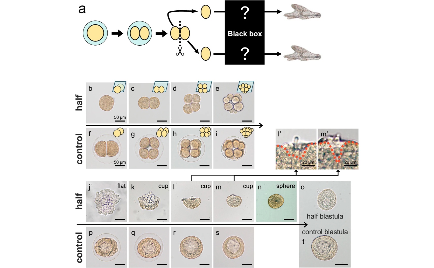
Contrary to the standard development of intact embryos (f–i, p–t), isolated blastomeres of the sea urchin (Hemicentrotus pulcherrimus) (a) undergo a unique self-organizing process involving distinct flat, cup, and sphere stages (b–e, j–o) to form a complete blastula.

Image adapted from: Suzuki et al. / CC BY (Source)

Key Findings

  • Sea urchin embryos can develop into complete organisms even when physically divided at the 2-cell stage
  • Isolated cells initially flatten but then reorganize into a spherical shape resembling a normal embryo
  • Proper body axis formation is re-established in these divided embryos through the Wnt/β-catenin signaling pathway
Animal embryos possess a remarkable ability to self-organize and develop into complete organisms, even when physically separated into individual cells early in development. This phenomenon, first demonstrated by Hans Driesch over a century ago, highlights the inherent robustness of developmental processes, yet the underlying molecular mechanisms remain largely unknown. Researchers at the University of Tsukuba[1] have now revisited these classic experiments using the sea urchin Hemicentrotus pulcherrimus to shed light on how divided embryos coordinate their development. The study focused on blastomeres – early embryonic cells – isolated from 2-cell stage H. pulcherrimus embryos. Unlike embryos allowed to develop normally, these isolated cells initially formed a flattened sheet of dividing cells before eventually rounding up into structures resembling blastulae. This difference in development suggested that the normal cues guiding embryonic organization were absent when cells were isolated. To understand what drives this altered development, the researchers employed live imaging and a technique called knockdown experiments. Knockdown experiments involve reducing the activity of specific genes to observe the effect on development. They found that activity of actomyosin – a protein complex responsible for cell contraction and movement – at the basal side of the cells, and structures called septate junctions, were crucial for the cells to round up. Septate junctions are cell-cell junctions that regulate the passage of molecules between cells and maintain the integrity of tissues[2]. Interestingly, the researchers observed that the normal anterior-posterior (A-P) and dorsal-ventral (D-V) axes of the embryo – essentially its head-to-tail and back-to-belly orientations – became disorganized during the initial stages of development. The original A-P poles of the isolated blastomeres came into contact as they formed a sphere. However, the embryos didn’t simply stop there. They actively corrected this disrupted axis, utilizing the well-known Wnt/β-catenin signaling pathway. This pathway is a key regulator of embryonic development in sea urchins and other animals, responsible for establishing body axes and cell fate. This finding is particularly significant because it demonstrates that divided embryos can re-establish proper body organization using pre-existing developmental mechanisms. The research builds upon earlier genomic work on H. pulcherrimus[3], which provided a detailed understanding of the sea urchin’s genome and created a database (HpBase) for researchers to easily access gene information and experimental protocols. This genomic resource was essential for identifying and studying the genes involved in axis formation and septate junction function. Furthermore, the study connects with research establishing H. pulcherrimus as a model organism for genome editing[4]. While sea urchins traditionally have been difficult to use for genetic studies due to their long breeding cycle, the development of genome editing tools in H. pulcherrimus paved the way for precise manipulation of genes like those involved in actomyosin activity and septate junction formation, enabling the knockdown experiments used in this study. The findings suggest that the ability to re-organize the A-P axis is essential for the successful development of divided embryos. The embryos aren’t simply relying on initial cell states, but actively working to correct any disruptions in body organization, highlighting the robustness of early embryonic development.

GeneticsMarine BiologyEvolution

References

Main Study

1) Unraveling the regulative development and molecular mechanisms of identical sea urchin twins

Published 5th September, 2025

https://doi.org/10.1038/s41467-025-63111-z


Related Studies

2) Identification of the genes encoding candidate septate junction components expressed during early development of the sea urchin, Strongylocentrotus purpuratus, and evidence of a role for Mesh in the formation of the gut barrier.

https://doi.org/10.1016/j.ydbio.2022.12.007


3) HpBase: A genome database of a sea urchin, Hemicentrotus pulcherrimus.

https://doi.org/10.1111/dgd.12429


4) TrBase: A genome and transcriptome database of Temnopleurus reevesii.

https://doi.org/10.1111/dgd.12780


Related Articles

An unhandled error has occurred. Reload 🗙