Deep Learning Tool Separates Plant Cells in 3D X-ray Images

Jenn Hoskins
21st January, 2024

Deep Learning Tool Separates Plant Cells in 3D X-ray Images

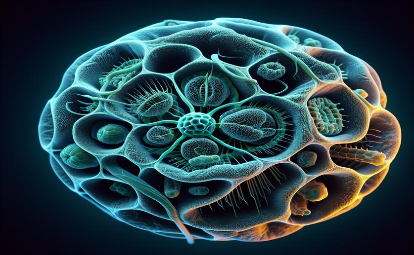
Stone cell clusters in pear tissue. (Top) 2D slices of contrast-enhanced micro-CT images of pear tissue samples of the inner cortex with stone cell clusters indicated with arrows. (Bottom) 3D visualization of the stone cell clusters

Image adapted from: Doorselaer et al. / CC BY (Source)
Understanding how plants function at a microscopic level is crucial for improving crop yields and resilience. The internal structure of plant tissues – how cells are arranged and the spaces between them – significantly impacts processes like gas exchange and water transport. Visualizing this structure in 3D is challenging, but recent advances in X-ray micro-computed tomography (micro-CT) offer a promising solution. However, a key limitation has been the difficulty in clearly identifying individual cells within the scans due to subtle differences in density between cells[1]. Researchers at KU Leuven have addressed this challenge with a new approach using artificial intelligence. The study focused on apple and pear fruit tissue, utilizing X-ray micro-CT to capture detailed 3D images. The core problem was that while micro-CT excels at showing the spaces between cells (the pore space), it struggles to define the cells themselves. This is because cells have similar densities, making it hard for the software to distinguish their boundaries. To overcome this, the researchers employed a ‘deep learning’ model – a type of artificial intelligence – to automatically identify and outline individual cells within the micro-CT scans. Deep learning models are trained by showing them many examples, and in this case, the model was trained on images of apple and pear tissue. The model learns to recognize patterns and features that define cell boundaries, even when those boundaries aren’t clearly visible due to density differences. The performance of the model was measured using a metric called the Aggregated Jaccard Index (AJI), which assesses the overlap between the model’s cell outlines and the actual cell boundaries. The best model achieved an AJI of 0.86 for apple tissue and 0.73 for pear tissue, representing a significant improvement over existing methods. This research builds upon earlier work demonstrating the importance of tissue microstructure in plant function. For example, studies have shown that the arrangement of cells and air spaces directly affects gas exchange, particularly oxygen and carbon dioxide transport[2]. Understanding these pathways is vital, as respiration in plant organs is heavily dependent on oxygen availability, often following a predictable pattern[2]. The new method developed by the KU Leuven team provides a way to accurately quantify the 3D structure of plant tissues, allowing for more detailed analysis of these gas exchange processes. Interestingly, the study found that apple tissue was easier to segment than pear tissue. This difference was linked to the porosity (amount of space) and surface area of the pore space within the tissues. Apple tissue had higher porosity and lower surface area, providing clearer ‘marker points’ for the AI to identify cell outlines. Conversely, pear tissue, with its lower porosity and higher surface area, presented a greater challenge. The presence of stone cells (brachysclereids) in pear tissue also complicated the segmentation process, as these structures influenced the arrangement of surrounding cells. This aligns with previous research highlighting how the geometry of cells and air spaces is a critical factor in gas exchange[2]. Furthermore, the study revealed that tissues with poor pore network connectivity were particularly difficult to segment. This suggests that a well-connected network of air spaces is not only important for gas transport but also for providing clear structural features that the AI can use for cell identification. Previous work using X-ray CT has focused on quantifying these void networks and their impact on oxygen diffusion[3], and this new method provides a complementary approach by allowing for detailed analysis of the cells themselves. The automated method developed in this study offers a significant advantage over traditional methods, which rely on time-consuming manual annotation or less accurate segmentation techniques. It allows researchers to rapidly and objectively quantify 3D cell morphology, which is crucial for understanding plant physiology and for studies where tissue anatomy plays a role, such as phenotyping and genotyping[3]. However, the researchers also caution that if tissue porosity is too low or the pore surface area too high, alternative scanning methods that enhance contrast may be necessary to obtain clear cell outlines.

FruitsBiotechPlant Science

References

Main Study

1) Automatic 3D cell segmentation of fruit parenchyma tissue from X-ray micro CT images using deep learning.

Published 19th January, 2024

https://doi.org/10.1186/s13007-024-01137-y


Related Studies

2) A three-dimensional multiscale model for gas exchange in fruit.

https://doi.org/10.1104/pp.110.169391


3) Automatic analysis of the 3-D microstructure of fruit parenchyma tissue using X-ray micro-CT explains differences in aeration.

https://doi.org/10.1186/s12870-015-0650-y


Related Articles

An unhandled error has occurred. Reload 🗙