Catnip-Silver Nanoparticles Show Superior Wound-Healing in Live Tissue Studies
Jim Crocker
13th June, 2024


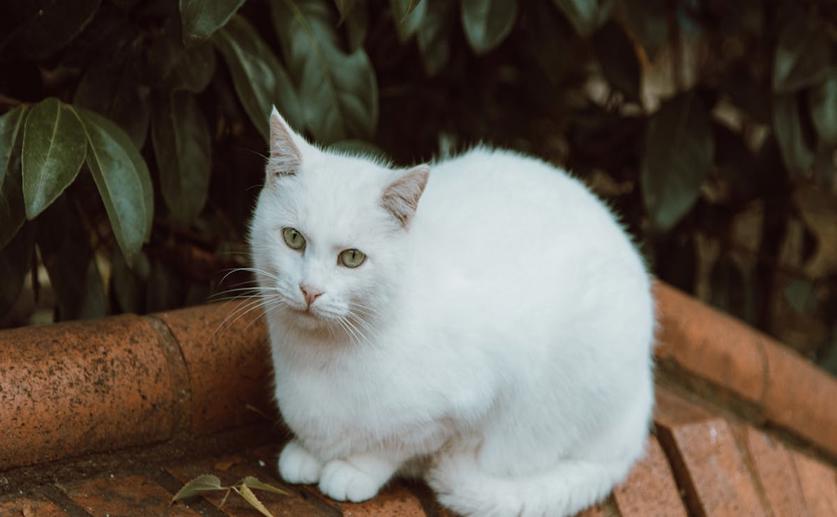
Catnip (Nepeta cataria)
Photographer: Dan Wilder
Key Findings
- Researchers at Burdur Mehmet Akif Ersoy University successfully synthesized silver nanoparticles (Nc-AgNPs) using Nepeta cataria (catnip) plant extract
- The Nc-AgNPs showed strong antibacterial properties, effectively inhibiting the growth of various bacterial strains, including E. coli, E. faecalis, and S. aureus
- In wound-healing tests on rats, the Nc-AgNPs combined with Vaseline significantly accelerated wound closure, achieving a 94% closure rate in 10 days, comparable to commercial treatments
References
Main Study
1) Superior In Vivo Wound-Healing Activity of Biosynthesized Silver Nanoparticles with Nepeta cataria (Catnip) on Excision Wound Model in Rat
Published 12th June, 2024
https://doi.org/10.1007/s12011-024-04268-4
Related Studies
2) Therapeutic Nanoparticles and Their Targeted Delivery Applications.
3) Cell Membrane Coating Nanotechnology.
4) Safe Nanoparticles: Are We There Yet?
Related Articles
 14th May, 2024 | Greg Howard