Deep Learning Tool Separates Plant Cells in 3D X-ray Images

Jenn Hoskins
21st January, 2024

Deep Learning Tool Separates Plant Cells in 3D X-ray Images

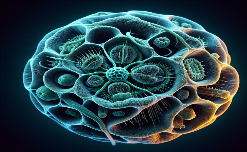
Image Source: Natural Science News, 2024

In the meticulous world of plant physiology, researchers dive deep into the microscopic universe of plant tissues. The ability to peek into this minute world, seeing the organization of cells and the tiny spaces between them, is crucial for understanding the physiological processes at work within a variety of plants. This understanding opens doors to insights that can go beyond pure science and influence agriculture, ecology, and our broader relationship with the natural world. Recently, an innovative approach using X-ray micro-computed tomography, or micro-CT, has emerged as a game-changer in plant research. Unlike the medical CT scans we're familiar with, micro-CT can reveal detailed 3D structures on a much smaller scale – think of it as a supercharged microscope that doesn't just magnify but unveils a rich three-dimensional understanding of the plant material it investigates. One fascinating application of this technology is in studying parenchyma, a type of tissue found in fruits like apples and pears. This tissue is full of cells and pockets of air (pore space) that all play their part in the fruit's development and ultimate ripeness. But there's a catch. Using conventional micro-CT techniques, pinning down the shape of each individual cell in the tissue can be a bit like trying to decipher a whisper in a windstorm. The cells just don't stand out enough from one another for the scanner to capture precisely where one ends and the next begins. But, behold! A leap forward has been achieved by researchers employing the power of deep learning – a remarkable type of artificial intelligence that learns and improves from experience much like a human brain might. This AI isn't playing chess or driving cars, though; it's learning to identify individual cells in fruit tissue images captured by micro-CT. In this recent study conducted by experts at KU Leuven in Belgium, deep learning-based models were meticulously trained to recognize the elusive outlines of cells in micro-CT images. The results are a notable step up from previous methods. The best deep learning model achieved an Aggregated Jaccard Index (a measure of accuracy in these segmentations) of 0.86 for apple tissue, surpassing the previous benchmark of 0.73. For pear tissue, the improvement was also significant, jumping from 0.67 to 0.73. And it's not just about cells. The AI was savvy enough to point out other key plant tissue structures, such as the ropes of vascular tissue that shuttle fluids around the plant, and clusters of tough cells known as brachysclereids, which are particularly influential in shaping pear tissue. One could argue that the art of distinguishing and segmenting apple cells from one another might be easier than doing the same with pear cells. This ease could be attributed to differences in the structure of the two fruits' tissues. Apples have more air space (porosity) among their cells and a simpler arrangement of that space, making it easier for the micro-CT scans to pick out individual cells. However, when the tissue has a lot of connectivity within the pore network or a high specific surface area (a more complex texture), even the best scans can flounder. In these situations, simply relying on a standard micro-CT scan might not be enough, and more sophisticated, contrast-enhanced scanning protocols might be necessary to discern the cell outlines properly. This study has paved the way for more refined quantification of 3D cell morphology in plant tissue, which traditionally would have required slow and painstaking manual work or less precise segmentation approaches. The researchers have gifted us with not just a snapshot but a detailed map of the minute and complex landscapes within our fruits. Now, while this might seem like a technical triumph reserved for scientists in lab coats, it has real-world implications. With the ability to understand and quantify plant tissue at this level, we can potentially boost the agricultural yield, improve our understanding of plant biology, and even work towards developing crops that are more resilient to diseases and the changing climate. Personally, I find it rather exciting that such tiny cells in fruits we enjoy on our kitchen tables can unlock such big potential. So next time you bite into a crisp apple or a juicy pear, remember, the world hidden within those bites is profoundly intricate and now, thanks to the fusion of technology and biology, a little less mysterious.

FruitsBiotechPlant Science

References

Main Study

1) Automatic 3D cell segmentation of fruit parenchyma tissue from X-ray micro CT images using deep learning.

Published 19th January, 2024

https://doi.org/10.1186/s13007-024-01137-y


Related Articles

An unhandled error has occurred. Reload 🗙